Robert J. Vissers
Aaron E Bair
Introduction
Surgical airway management is defined as the creation of an airway by an invasive technique. All other methods of airway management use existing anatomical portals of access to the trachea (i.e., nasopharynx, oropharynx). Surgical airway management involves the creation of an opening to the trachea by surgical means. This opening is then used to provide ventilation and oxygenation. There is some confusion engendered by use of the term surgical airway management. In some discussions, surgical airway management includes both cricothyrotomy and needle cricothyrotomy with percutaneous transtracheal ventilation (PTV). Other discussions limit surgical airway management to cricothyrotomy, and consider PTV to be simply another airway management technique. For the purposes of discussion in this chapter, surgical airway management is deemed to include cricothyrotomy, PTV, and placement of a surgical airway using a cricothyrotome, which is a device intended to place a surgical airway percutaneously, usually in one or two steps, without performance of formal cricothyrotomy. The cricothyrotomes may be considered as an alternative to cricothyrotomy; however, cricothyrotomes that place an uncuffed tube do not protect the airway.
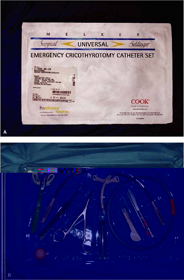
PTV through a catheter is rarely, if ever, done in adults. It does not protect the airway and is grossly inferior to cricothyrotomy in terms of both airway protection and gas exchange. In adults, cricothyrotomy by open or Seldinger technique is preferred. PTV should be reserved for children younger than 12 years, whose anatomy is not conducive to cricothyrotomy. The faculty of The Difficult Airway Course: Emergency recently designed a kit that offers the instruments and equipment to perform either the Seldinger percutaneous cricothyrotomy or an open cricothyrotomy, using a cuffed tube for both methods (Melker Universal Emergency Cricothyrotomy Catheter Set, Cook Critical Care, Bloomington, IN; Fig. 16-1). Each main surgical airway technique is described in detail in the sections that follow.
Description
Cricothyrotomy is the establishment of a surgical opening in the airway through the cricothyroid membrane and placement of a cuffed tracheostomy tube or endotracheal tube (ETT).
A cricothyrotome is a kit or device that is intended to establish a surgical airway without resorting to formal cricothyrotomy. These kits use two basic approaches. One approach relies on the Seldinger technique, in which the airway is accessed via a small needle through which a flexible guide wire is passed. The airway device, with a dilator, is then passed over this guide wire and into the airway in a manner analogous with that of central line placement by the Seldinger technique. The other technique relies on the direct percutaneous placement of an airway device without the use of a Seldinger technique. There have been no clinical studies to date demonstrating the superiority of one approach over another or of any of these devices over formal surgical cricothyrotomy. However, certain attributes of the devices make them intuitively more, or less, hazardous for insertion (see the Evidence section).
Indications and Contraindications
The primary indication for cricothyrotomy is when a failed airway has occurred (see Chapter 2) and the patient cannot be adequately ventilated or oxygenated with a bag and mask, or the patient is adequately oxygenated, but there is not another available device (e.g., fiberoptic scope, lighted stylet, intubating laryngeal mask airway [LMA]) that is believed likely to successfully secure the airway. A second indication is a method of primary airway management in patients for whom intubation is contraindicated or believed to be impossible. Thus, cricothyrotomy should be thought of as a rescue technique in most circumstances, and only infrequently will it be used as the primary method of airway management. An example of a circumstance in which cricothyrotomy is the primary method of airway management is the patient with severe lower facial trauma in whom access through the mouth or nose would be too time consuming or impossible. This patient requires immediate airway management because of the risk of aspiration of blood and secretions, and cricothyrotomy is indicated.
|
|
|
Figure 16-1 • A: Melker Universal Emergency Cricothyrotomy Catheter Set (Cook Critical Care, Bloomington, IN). B: Opened set containing cuffed tracheostomy tube, as well as equipment for both open surgical and Seldinger techniques. |
The primary hurdle to performing cricothyrotomy is simply recognizing when it is necessary to proceed with surgical airway management, and abandoning further attempts at laryngoscopy, or the use of an alternative device. Rapid sequence intubation (RSI), augmented by a variety of noninvasive airway management methods, is so successful that cricothyrotomy is often viewed as a method of last resort, to be undertaken only after multiple noninvasive attempts or techniques have failed. However, the relentless, unsuccessful, pursuit of a noninvasive airway, along with the resultant delay in the initiation of a surgical airway, can readily result in hypoxic disaster. This fact is particularly true in the “can't intubate, can't oxygenate” (CICO) circumstance, when surgical airway management is immediately indicated and must not be delayed for attempts using other devices.
Once the decision to initiate surgical airway management has been made, there are a few fundamental considerations:
a. Will accessing the cricothyroid membrane be effective? In other words, will an incision at the level of the cricothyroid membrane bypass the obstruction and solve the problem? If the obstructing lesion is significantly distal to the cricothyroid membrane, performing a cricothyrotomy is a critical waste of time (see Chapter 36).
b. Will the patient's anatomy or pathological process make cricothyrotomy difficult to perform? Placement of the initial skin incision is based on palpating the pertinent anatomy. If adiposity, burns, trauma, or infection make this procedure difficult, then the strategy should be adjusted accordingly. A mnemonic for difficult cricothyrotomy (SHORT) is shown in Box 16-1 and is discussed in Chapter 7.
c. Which type of invasive technique will most readily be used (i.e., open surgical or percutaneous)? This consideration takes into account provider preference based on previous experience and equipment availability.
Contraindications to surgical airway management are few and, with one exception, are relative. That one exception is young age. Children have a small, pliable, mobile larynx and cricoid cartilage, making cricothyrotomy extremely difficult. For children younger than 12 years, unless they are teenage or adult size, PTV should be used as the surgical airway management technique of choice (see Chapters 21 and 22). Relative contraindications include pre-existing laryngeal or tracheal pathology such as tumor, infections, or abscess in the area in which the procedure will be performed; hematoma or other anatomical destruction of the landmarks that would render the procedure difficult or impossible; coagulopathy; and lack of operator expertise. Cricothyrotomy has been performed successfully after systemic thrombolytic therapy. Cricothyrotomy has a high success rate when performed in the emergency department (ED) setting. The presence of an anatomical barrier, in particular, should prompt consideration of alternative techniques that might result in a successful airway. However, in cases in which no alternative method of airway management is likely to be successful or timely enough, cricothyrotomy should be performed without hesitation. The same principles apply for both the cricothyrotome and for PTV.
BOX 16-1 SHORT Mnemonic for Difficult Cricothyrotomy
· Surgery (history of neck surgery, presence of surgical scar)
· Hematoma
· Obesity
· Radiation (history or evidence of radiation therapy)
· Trauma (direct laryngeal trauma with disrupted landmarks)
PTV is not contraindicated in small children and, in fact, is the surgical airway method of choice for children younger than 12 years. The cricothyrotomes have not been demonstrated to improve success rates or time or to decrease complication rates when compared with surgical cricothyrotomy. As with formal cricothyrotomy, experience, skill, knowledge of anatomy, and adherence to proper technique are essential for success when a cricothyrotome is used.
Technique
Anatomy and Landmarks
The cricothyroid membrane is the anatomical site of access in the emergent surgical airway, regardless of the technique used. It has several advantages over the trachea in the emergent setting. The cricothyroid membrane is more anterior than the lower trachea, and there is less soft tissue between the membrane and the skin. There is less vascularity and less chance of significant bleeding.
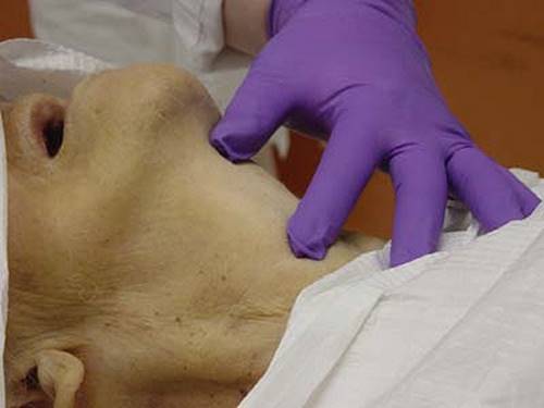
The cricothyroid membrane is identified by first locating the laryngeal prominence (notch) of the thyroid cartilage. Approximately one fingerbreadth below the laryngeal prominence, the membrane may be palpated in the midline of the anterior neck, as a soft depression between the inferior aspect of the thyroid cartilage above and the hard cricoid ring below. The relevant anatomy may be easier to appreciate in males because of the more prominent thyroid notch. The thyrohyoid space, which lies above the laryngeal prominence, and the hyoid bone, which resides high in the neck, should also be identified. This will prevent the misidentification of the thyrohyoid membrane as the cricothyroid membrane, which would lead to misplacement of the tracheostomy tube above the vocal cords. In children, the cricothyroid membrane is disproportionately smaller because of a greater overlap of the thyroid cartilage over the cricoid cartilage. For this reason, cricothyrotomy is not recommended in children age 12 years or younger.
Unfortunately, the same anatomical or physiological abnormalities (i.e., trauma, morbid obesity, congenital anomalies) that necessitated the surgical airway may also hinder easy palpation of landmarks. One way of estimating the location of the cricothyroid membrane is by placing four fingers on the neck, oriented longitudinally, with the small finger in the sternal notch. The membrane is approximately located under the index finger and can serve as a point at which the initial incision is made. Except as described later in the technique for the rapid four-step cricothyrotomy, a vertical skin incision is preferred, and particularly so if anatomical landmarks are not readily apparent. Palpation through this vertical incision can then confirm the location of the cricothyroid membrane. Alternatively, identification may be assisted by using a locator needle, attached to a syringe containing saline or lidocaine. Aspiration of air bubbles suggests entry into the airway, but it will not distinguish between the cricothyroid membrane and a lower tracheal placement.
The No-drop Technique
The cricothyrotomy instrument set should be simple, consisting of only the equipment necessary to complete the procedure. A sample listing of recommended contents of a cricothyrotomy tray is shown in Box 16-2. Commercial kits are now available that also contain the instruments required for a cricothyrotomy (Fig. 16-1).
1. Identify the landmarks. The cricothyroid membrane is identified using the landmarks described previously (Fig. 16-2).
2. Prepare the neck. If time permits, apply appropriate antiseptic solution. Local anesthesia is desirable if the patient is conscious. Infiltration of the skin and subcutaneous tissue of the anterior neck with 1% lidocaine solution will provide adequate anesthesia. If time permits and the patient is conscious and responsive, anesthetize the airway by injecting lidocaine by transcricothyroid membrane puncture (see Chapter 8). The patient will cough briefly, but the airway will be reasonably anesthetized and further cough reflexes suppressed.
3. Immobilize the larynx. Throughout the procedure, the larynx must be immobilized (Fig. 16-3). This is best done by placing the thumb and long finger on opposite sides of the superior laryngeal horns, the posterior superior aspect of the laryngeal cartilage. With the thumb and long finger thus placed, the index finger is ideally positioned anteriorly to relocate and reidentify the cricothyroid membrane at any time during the procedure.
4. Incise the skin. A 2-cm vertical midline skin incision should be used (Fig. 16-4). Care should be taken to avoid cutting the deeper structures of the neck. The cricothyroid membrane is separated from the outside world only by skin, subcutaneous tissue, and anterior cervical fascia. An overly vigorous incision risks damage to the larynx, cricoid cartilage, and the trachea.
|
|
|
Figure 16-2 • Anatomy of the Larynx. The cricothyroid membrane (arrow) is bordered above by the thyroid cartilage and below by the cricoid cartilage. |
|
|
|
Figure 16-3 • A: Surface anatomy of the airway. B: The thumb and long finger immobilize the superior cornua of the larynx; the index finger is used to palpate the cricothyroid membrane. |
5. Reidentify the membrane. With the thumb and long finger maintaining immobilization of the larynx, the index finger can now palpate the anterior larynx, the cricothyroid membrane, and the cricoid cartilage without any interposed skin or subcutaneous tissue (Fig. 16-5). The landmarks thus confirmed, the index finger can be left in the wound by placing it on the inferior aspect of the anterior larynx, thus providing a clear indicator of the superior extent of the cricothyroid membrane.
6. Incise the membrane. The cricothyroid membrane should be incised in a horizontal direction, with an incision at least 1 cm long (Fig. 16-6A). It is recommended to try to incise the lower half of the membrane rather than the upper half because of the relatively cephalad location of the superior cricothyroid artery and vein; however, this may be unrealistic in the emergent setting (Fig. 16-6B).
|
|
|
Figure 16-4 • With the index finger moved to the side but continued firm immobilization of the larynx, a vertical midline skin incision is made, down to the depth of the laryngeal structures. |
|
|
|
Figure 16-5 • With skin incised, the index finger can now directly palpate the cricothyroid membrane. |
|
|
|
Figure 16-6 • A: A horizontal membrane incision is made near the inferior edge of the cricothyroid membrane. The index finger may be swung aside or may remain in the wound, palpating the inferior edge of the thyroid cartilage, to guide the scalpel to the membrane. B: A low cricothyroid incision avoids the superior cricothyroid vessels, which run transversely near the top of the membrane. |
7. Insert the tracheal hook. The tracheal hook is rotated so that it is oriented in the transverse plane, passed through the incision, and then rotated again so the hook is oriented in a cephalad direction. The hook is then applied to the inferior aspect of the thyroid cartilage, and gentle upward and cephalad traction is applied to bring the airway immediately out to the skin incision (Fig. 16-7). If an assistant is available, this hook may be passed to the assistant to maintain immobilization of the larynx.
8. Insert the Trousseau dilator. The Trousseau dilator may be inserted in one of two ways. One method is to insert the dilator well in through the incision, directing the blades of the dilator longitudinally down the airway. The second method, which is preferred, is to insert the dilator minimally into the anterior wound with the blades oriented superiorly and inferiorly, allowing the dilator to open and enlarge the vertical extent of the cricothyroid membrane incision, which is often the anatomically limiting dimension (Fig. 16-8). When this technique is used, care must be taken not to insert the dilator too deeply into the airway because it will impede subsequent passage of the tracheostomy tube.
|
|
|
Figure 16-7 • A: The tracheal hook is oriented transversely during insertion. B and C: After insertion, cephalad traction is applied to the inferior margin of the thyroid cartilage. |
9. Insert the tracheostomy tube. The tracheostomy tube, with its inner cannula in situ, is gently inserted through the incision between the blades of the Trousseau dilator. As the tube is advanced gently following its natural curve, the Trousseau dilator is rotated to allow the blades to orient longitudinally in the airway (Fig. 16-9). The tracheostomy tube is advanced until it is firmly seated against the anterior neck. The Trousseau dilator is then carefully removed.
10. Inflate the cuff, and confirm tube position. With the cuff inflated, the tracheostomy tube position can be confirmed by the same methods as the ETT position. Carbon dioxide (CO2) detection will reliably indicate correct placement of the tube and is mandatory, as for endotracheal intubation. Immediate subcutaneous emphysema with bagging suggests probable paratracheal placement. If doubt remains, rapid insertion of a nasogastric tube through the tracheostomy tube will result in easy passage if the tube is in the trachea and obstruction if the tube has been placed through a false passage into the tissues of the neck. Auscultation of both lungs and the epigastric area is also recommended, although esophageal placement of the tracheostomy tube is exceedingly unlikely. Chest radiography should be performed to assist in the assessment of tube placement and to evaluate for the presence of barotrauma.
|
|
|
Figure 16-8 • A: The Trousseau dilator is inserted a short distance into the incision. B: In this orientation, the dilator enlarges the opening vertically, the crucial dimension. |
BOX 16-2 Recommended Contents of Cricothyrotomy Tray
· Trousseau dilator
· Tracheal hook
· Scalpel with no. 11 blade
· Cuffed, nonfenestrated, no. 4 tracheostomy tube
· Optional equipment: several 4 × 4 gauze sponges, two small hemostats, and surgical drapes
The Rapid Four-step Technique
This abbreviated cricothyrotomy method has been developed and adopted for training purposes at some centers. As with all techniques, the patient should be maximally oxygenated and, if given sufficient time, the anterior neck may be prepared and locally anesthetized as for the no-drop method. From a position at the head of the bed, the rapid four-step technique for cricothyrotomy proceeds sequentially:
|
|
|
Figure 16-9 • A: Insertion of the tracheostomy tube. B: Rotation of the Trousseau dilator to orient the blades longitudinally in the airway facilitates passage of the tracheostomy tube. C: Tracheostomy tube fully inserted; instruments removed. |
|
|
|
Figure 16-10 • Palpation: the operator's thumb is on the hyoid bone, while the cricothyroid membrane is identified using the index finger. |
1. Palpate and identify landmarks. The cricothyroid membrane should be identified as described previously (Fig. 16-10). If the key landmarks are unable to be identified by palpation through the soft tissue, then a vertical skin incision is required to permit accurate identification.
2. Make skin incision. Once the pertinent palpable anatomy is identified, the cricothyroid membrane is incised. If the anatomy is fully appreciated through the intact skin, and there is no uncertainty about landmarks or location, then incise the skin and cricothyroid membrane simultaneously with a single horizontal incision of approximately 1.5 cm in length (Fig. 16-11A). For this type of incision, a no. 20 scalpel yields an incision that requires little widening, once used to puncture the skin and cricothyroid membrane. If the anatomy is not readily and unambiguously identified through the skin, then an initial vertical incision should be created to allow more precise palpation of the anatomy and identification of the cricothyroid membrane. In either situation, the cricothyroid membrane is incised with the no. 20 blade that is maintained in the airway, while a tracheal hook (preferably a blunt hook) is placed parallel to the scalpel on the caudad side of the blade (Fig. 16-11B). The hook is then rotated to orient it in a caudad direction to put gentle traction on the cricoid ring. The scalpel is then removed from the airway. At no time during this procedure is the incision left without instrument control of the airway. This detail is particularly important in a scenario where the patient still has the ability to respond or swallow. The newly created stoma could be irretrievably lost if the airway is uncontrolled and moves relative to the skin incision. In addition, as is the case for the no-drop method, this is a technique that relies exclusively on palpation of key structures. Bleeding will inevitably obscure visualization of the anatomy. No time should be wasted using suction or gauze or manipulating the overhead lighting.
3. Apply traction. The tracheal hook that has been rotated caudally and is controlling the cricoid ring is now used to lift the airway toward the skin incision. This action provides modest stoma dilation. The direction of hook pull is reminiscent of the up and away direction used with laryngoscopy (Fig. 16-12). The amount of traction force required for easy intubation (18 newtons or 4.05 pounds force) is significantly lower than the force that is associated with breakage of the cricoid ring (54 newtons or 12.14 pounds force). Use of the hook in this direction generally provides sufficient widening of the incision, and a Trousseau dilator is usually not required. The technique of pulling the airway upward in this way also minimizes the possibility of intubating the pretracheal potential space.
|
|
|
Figure 16-11 • Incision: A: A horizontal incision is initiated while stabilizing the larynx. B: Before removing the scalpel from the airway, a hook is placed on the caudal side of the scalpel, parallel to the blade. |
4. Intubate. With adequate control of the airway using the hook placed on the cricoid ring, tracheostomy tube is readily placed into the airway and secured (Fig. 16-13). Confirmation techniques proceed as described in the no-drop technique.
Complications
Because of the common adoption of RSI, cricothyrotomy is infrequently performed in EDs, so reports of complications are difficult to evaluate. In the National Emergency Airway Registry (NEAR II) study, less than 1% of more than 7,700 ED intubations involved cricothyrotomy.
|
|
|
Figure 16-12 • Traction: the hook is applied to the cricoid ring and lifted. |
The most important complication for the patient in the context of surgical airway management is when delayed decision making after initial intubation failure leads to prolonged, ineffective intubation attempts that result in hypoxic injury. Failure to rapidly place the tracheotomy tube into the trachea or misplacement of the tube into the soft tissues of the neck is more a failure of technique than a complication and must be recognized immediately, as is the case with any misplaced ETT. Complications such as pneumothorax, significant hemorrhage requiring operative intervention, laryngeal or tracheal injury, and long-term complications, such as subglottic stenosis or permanent voice change, are relatively infrequent and usually minor. The potential for these complications to occur in no way outweighs the need to establish the airway. In general, the incidence of all complications, immediate and delayed, and major or minor, is approximately 20%. Most of these complications are minor, particularly when compared to the consequences of a persistently failed airway. Box 16-3 lists complications of surgical airway management.
|
|
|
Figure 16-13 • Intubation: the tracheostomy tube is passed into the incision as the hook stabilizes the cricoid ring. |
BOX 16-3 Complications of Surgical Airway Management
· Hemorrhage
· Pneumomediastinum
· Laryngeal/tracheal injury
· Cricoid ring laceration
· Barotrauma (transtracheal jet ventilation)
· Infection
· Voice change
· Subglottic stenosis
Cricothyrotome Technique
Seldinger Technique
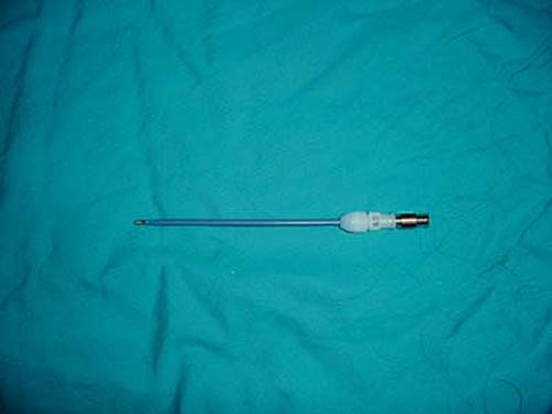
Numerous commercial cricothyrotome devices are available. The Melker Universal Emergency Cricothyrotomy Catheter Set uses a modified Seldinger technique to assist in the placement of a tracheal airway (Figs. 16-1 and 16-14A). This method is similar to the one commonly used in the placement of central venous catheters and offers some familiarity to the operator uncomfortable with or inexperienced in the surgical cricothyrotomy technique described previously. Devices that incorporate an inflatable cuff are recommended (Fig. 16-14B).
1. Identify landmarks. The cricothyroid membrane is identified by the method described previously. The nondominant hand is used to control the larynx and maintain identification of the landmarks.
2. Prepare neck. Antiseptic solution is applied to the anterior neck, and, if time permits, infiltration of the site with 1% lidocaine with epinephrine is recommended.
3. Insert locator needle. The locator needle (18-gauge) is then inserted into the cricothyroid membrane in a slightly caudal direction (Fig. 16-14C). The needle is attached to a syringe and advanced with the dominant hand, while negative pressure is maintained on the syringe. The sudden aspiration of air indicates placement of the needle into the tracheal lumen.
4. Insert guide wire. The syringe is then removed from the needle. A soft-tipped guide wire is inserted through the needle into the trachea in a caudal direction (Fig. 16-14D). The needle is then removed, leaving the wire in place. Control of the wire must be maintained at all times.
5. Incise skin. A small skin incision is then made adjacent to the wire. This facilitates passage of the airway device through the skin (Fig. 16-14E). Alternatively, the skin incision may be made vertically over the membrane before insertion of the needle and guide wire.
6. Insert the airway and dilator. The airway catheter (3–6 mm internal diameter [ID]) with an internal dilator in place is inserted over the wire into the trachea (Fig. 16-14F). If resistance is met, the skin incision should be deepened and a gentle twisting motion applied to the airway device (Fig. 16-14G). When the airway device is firmly seated in the trachea, the wire and dilator are removed together (Fig. 16-14H).
|
|
|
Figure 16-14 • A: Kit contents. B: Cuffed tube. C: Needle insertion. D: Wire placement through needle. E: Small incision. F: Airway with dilator inserted with wire guidance. G: Airway inserted to the hub using a gentle twisting motion. H: Wire and dilator removed as one. (Melker Universal Cricothyrotomy Kit, Cook Critical Care, Bloomington, IN.) |
7. Confirm tube location. If the device has a cuff, inflate it at this time. Tube location can then be confirmed as for surgical cricothyrotomy, including mandatory end-tidal CO2 detection. The devices are radiopaque on radiographs. The airway must then be secured properly.
Direct Airway Placement Devices
Several direct airway devices (e.g., Nu-Trake, Pertrach) are commercially available. These generally involve multiple steps in the insertion, using a large device that functions as both introducer and airway. The details of the operation of these devices may be obtained from the manufacturer and are provided as inserts with the kits. These devices offer no clear advantage in technique, are rarely (if ever) as easily placed as is claimed, and are considered more likely to cause traumatic complications during their insertion than those that use a Seldinger technique, primarily because of the cutting characteristics of the airway device. In particular, cricothyrotomes recommended for children should be approached with extreme caution and are not recommended.
Percutaneous Transtracheal Jet Ventilation Technique
Needle cricothyrotomy with percutaneous transtracheal jet ventilation (TTJV) is a surgical airway that may be used to temporize in the CICO situation, particularly in children. Although TTJV is rarely performed in the emergency setting, it is a simple, relatively effective means of supporting oxygenation. Advantages of this technique over cricothyrotomy may include speed, a simpler technique, and less bleeding. It can also provide an alternative for operators unable to perform a cricothyrotomy. Age is not a contraindication to TTJV, which is the surgical airway of choice for children younger than 12 years (see Chapters 20,21,22).
Several other aspects of this technique that differ from cricothyrotomy are important to consider. To provide ventilation, supraglottic patency must be maintained to allow for exhalation. In the case of complete upper-airway obstruction, air stacking from TTJV will cause barotrauma; therefore, cricothyrotomy is preferable. Another significant difference is that the catheter in TTJV does not provide airway protection. Also, suctioning cannot adequately be performed through the percutaneous catheter. TTJV has been associated with a significant incidence of barotrauma and is less commonly used as a rescue device, particularly with the widespread use of other devices such as the LMA. TTJV is therefore best considered a temporizing means of rescue oxygenation until a more definitive airway can be obtained.
a. Procedure
1. Identify the landmarks. The anatomy and landmarks used in needle cricothyrotomy are identical to those described previously for a surgical cricothyrotomy. If there are no contraindications, the head of the patient should be extended. Placing a towel under the shoulders may facilitate cervical hyperextension. The area overlying the cricothyroid membrane should be prepared with an antiseptic solution and, if time permits, anesthetized with 1% lidocaine and epinephrine.
2. Immobilize the larynx. Use the thumb and the middle fingers of the nondominant hand to stabilize the larynx and cricoid cartilage while the index finger palpates the cricothyroid membrane. It is essential to maintain control of the larynx throughout the procedure.
3. Insert transtracheal needle. A large-bore intravenous (IV) catheter (12–16 gauge) is attached to a 20-mL syringe, which may be empty or partially filled with a clear liquid. A 15-degree angle can be created by bending the needle/catheter combination 2.5 cm from the distal end of the IV catheter, or a commercially available catheter can be employed (Fig. 16-15A). A commercial catheter may be preferred because it is reinforced with wire coils to prevent kinking (Fig. 16-16). The dominant hand holds the syringe with the needle directed caudally in the long axis of the trachea at a 30-degree angle to the skin (Fig. 16-15B). While maintaining negative pressure on the syringe, the needle is inserted through the cricothyroid membrane into the trachea. As soon as the needle enters the trachea, the syringe will easily fill with air. If a liquid is used, bubbles will appear. Any resistance implies that the catheter remains in the tissue. In the awake patient, lidocaine may be used in the syringe and then injected into the tracheal lumen to suppress the cough reflex.
|
|
|
Figure 16-15 • A: Nonkinking catheter for jet ventilation. B: Needle insertion. C: Removal of needle. D: Tracheotomy catheter placed. E: Oxygenation with bag. F: Oxygenation with jet ventilator. (Acutronic, Germany). |
|
|
|
Figure 16-16 • Nonkinkable wire-coiled transtracheal jet ventilation catheter (Cook Critical Care, Bloomington, IN). |
4. Advance catheter. Once entry into the trachea is confirmed, the catheter can be advanced. The needle may be partially or completely withdrawn before advancement; however, the needle should not be advanced with the catheter. A small incision can assist with catheter advancement if there is resistance at the skin (Fig. 16-14C and D).
5. Confirm location. The catheter should be advanced to the hub and controlled by hand at all times. Air should be reaspirated to confirm once again the location of the catheter within the trachea.
6. Connect to bag or jet ventilation. The catheter may be connected to a bag ventilator using a pre-existing adapter, or if there is only a Luer lock present, the adapter from a 3-0 pediatric ETT will fit the Luer lock (Fig. 16-15E). For jet ventilation, the catheter is connected to the female end of the tubing of the jet ventilation system by a Luer lock. The hub should not be secured in place by anything other than a human hand until a definitive airway is established (Fig. 16-15F). Firm, constant pressure must be applied by hand to ensure that proper positioning is maintained and to create a seal at the skin to minimize air leak.
7. Perform jet ventilation. In the adult, the jet ventilation system should be connected to an oxygen source of 50 pounds per square inch (psi) with a continuously adjustable regulator to allow the pressure to be titrated so the lowest effective pressure (often about 30 psi) required to safely deliver a tidal volume is used. In general, inspiration is less than 1 second followed by 3 seconds of expiration. Because the gas flow through a 14-gauge needle at 50 psi is 1,600 mL/second, less than 1 second of inspiratory time is required for an adequate tidal volume in a normally compliant lung. Exhalation depends on the elastic recoil of the lung, which is a relatively low driving pressure. Therefore. the recommended inspiratory-to-expiratory ratio (I:E) is 1:3. It is important to maintain upper-airway patency to allow for exhalation and avoid air trapping and barotrauma. All patients should have an oral and nasal airway placed. For small adults and children, oxygen pressure should be downregulated to less than 20 to 30 psi, if possible. For children younger than 5 years, a bag should be used for ventilation, connected to the catheter using the ETT adapter from a 3.0-mm-ID ETT.
b. b. Equipment
1. Transtracheal catheters. A large-bore IV catheter is acceptable. The proper placement is made easier by placing a small angle 2.5 cm from the tip. Commercially available devices include precurved (Acutronic, Germany) and nonkinkable wire-coiled (Cook Critical Care, Bloomington, IN) catheters (Figs. 16-15A and 16-16). The wire-coiled catheter will not kink when bent; therefore, it provides a more secure airway.
2. Transtracheal jet ventilation systems. The TTJV system consists of a high-pressure oxygen source (usually central wall oxygen pressure of 50 psi), high-pressure oxygen tubing, a regulator to control the driving pressure, an on-off valve to control inspiratory time, high-pressure tubing, and a Luer lock to connect to the catheter (Fig. 16-17).
A regulator to control the driving pressure is optional but recommended. This device is particularly useful where barotrauma is a concern and in pediatrics, where the inspiratory pressures should be reduced to less than 20 to 30 psi, if possible. Although a system can be assembled inexpensively from readily available materials, a commercially made, preassembled system is recommended. The reliability and control inherent in the commercial devices are well worth the marginal increase in cost.
|
|
|
Figure 16-17 • Disposable jet ventilator system with high-pressure oxygen tubing, on-off valve, and PVC tubing with Luer lock. Note that this device does not include a pressure regulator (Cook Critical Care, Bloomington, IN). |
A TTJV system can also be connected to a low-flow portable oxygen tank when circumstances require mobility. When the flow is set at the maximal 15 L/minute and no flow is allowed, the pressure temporarily increases to 120 psi. Once flow is released, high flow occurs momentarily and then rapidly decreases to the steady state of 5 to 10 psi. Adequate tidal volumes may be achieved through a 14-gauge catheter in the first 0.5 second. A shorter I:E ratio of 1:1 is recommended.
Another setup using manual ventilation with a self-inflating reservoir bag has been described, using standard equipment found in any ED. Bag ventilation may be connected directly to the percutaneous transtracheal catheter in two ways. The male end of a 15-mm ETT adapter from a 3-mm-ID ETT will fit directly into the catheter. Alternatively, the male end of a plungerless 3-mL syringe will fit into the catheter, and the male end of an 8-mm-ID ETT adapter will then insert into the female end of the empty syringe. Ventilation is temporary at best, and partial arterial CO2pressure (PaCO2) will increase at a rate 2 to 4 mm Hg/minute. Even the simple assembly of this system is too time consuming to be done during the event, so it must be preassembled. This arrangement may have particular utility in the pediatric patient younger than 5 years when excessive pressures may be delivered via a TTJV device, even when a regulator to control inspiratory pressures is available. In general, children younger than 5 years should receive TTV via a ventilation bag; ages 5 to 12 years old at less than 30 mm Hg and older than 12 years to adult at 30 to 50 mm Hg. A catheter of less than 3 mm ID will be insufficient to adequately ventilate/oxygenate the adult patient using a bag, and 50 psi pressurized oxygen is required.
c. Complications specific to transtracheal jet ventilation
· Subcutaneous emphysema
· Barotrauma
· Reflex cough with each ventilation (may be aborted with lidocaine)
· Catheter kinking
· Obstruction from blood or mucus
· Esophageal puncture
· Mucosal damage if nonhumidified gas is used
Tips and Pearls
Surgical airway management is rarely the method of first choice for patients requiring emergency airway management. However, there is a population of patients for whom surgical airway management will literally make the difference between life and death. Therefore, emergency physicians and others who provide care for patients requiring emergency airway management must be proficient with surgical airway management.
There may be little advantage to using a cricothyrotome rather than a formal, surgical cricothyrotomy set. Time of performance of the procedure, complication rates, degree of difficulty, and success rates are all comparable between the two methods. Of the available cricothyrotomes, those that use the Seldinger technique and use a cuffed tube are preferable. Personal preference should also guide selection. A new kit offers all instruments and equipment to perform both the Seldinger-based cricothyrotome insertion and a formal, open, cricothyrotomy by either the standard “no-drop” or rapid four-step technique (Melker Universal Emergency Cricothyrotomy Catheter Set, Cook Critical Care, Bloomington, IN; Fig. 16-1). There is no evidence that any cricothyrotome can be placed in a child younger than 10 to 12 years of age with acceptable success and safety, regardless of the design of the device or the claims of the manufacturer.
PTV is virtually never indicated in the adult patient. In adults, establishment of a more functional surgical airway using a cricothyrotome or by formal cricothyroidotomy is vastly preferable. However, PTV remains a useful temporizing measure. In children younger than 12 years, the opposite is true. In this age group, PTV is the primary surgical airway management method of choice, and cricothyrotomy and cricothyrotomes should be avoided. Despite the extreme infrequency of use of PTV in the ED, it is important to have a PTV set readily available and to be familiar with how to connect and use it. The wire-coiled catheter designed for TTJV is preferable to standard IV catheters because of the tendency for the latter to kink.
Of the methods described in this chapter, only a formal surgical cricothyrotomy and variations of the recently modified Melker set result in the placement of a cuffed tube within the trachea. The other techniques described here must be considered temporary at best. Placement of a tracheostomy tube or ETT through a formal surgical cricothyrotomy incision results in an airway that can be used as a definitive airway for the patient.
The no. 4 cuffed tracheostomy tube, which has an inside diameter of 5 mm, should be used for virtually all cases of adult cricothyrotomy in the ED. The tube is of adequate size to provide ventilation in virtually all circumstances, and its outside dimensions are such that it will almost always be easily inserted. For very large adult men, a no. 6 can be used.
Evidence
1. Which technique is best? There is almost no literature that effectively compares different techniques of invasive surgical airway management. The relatively rare performance of an emergency surgical airway, compounded with the urgency of the circumstance, may explain the absence of any controlled clinical trials comparing techniques in the emergency setting. Comorbid injuries or illnesses often preclude long-term assessment of sequelae, and the few studies performed do not compare or identify specific invasive techniques (1). As such, the current level of evidence for or against a particular technique exists as expert consensus based on collective experience, limited descriptive series, or studies in cadaveric and animal models (2,3,4,5,6,7,8,9,10).
Several studies have directly compared the percutaneous, wire-guided technique to the traditional open, or no-drop, technique in cadavers (11,12,13). Eisenberger et al. (11), studying the performance of intensive care trainees, and Chan et al. (12), in a similar study of emergency residents and attendings, compared the two techniques in randomized, cross-over studies and found no difference in times to completion between groups. Immediate complications were also similar. Interestingly, the majority of participants in Chan's study stated a preference for the percutaneous, wire-guided technique (12).
2. Rapid four-step or no-drop technique for open cricothyrotomy? Each approach has its proponents. The no-drop technique has been in use for decades and has withstood the test of time. The rapid four-step technique (RFST) is proposed to be an improvement over the no-drop technique based on the following:
a. The rapid four-step technique requires only one person to perform the procedure. Ideally, the no-drop technique requires two people (i.e., one operator and one assistant). The assistant is responsible for not dropping the tracheal hook as it stabilizes the trachea. Without an assistant, the operator must maintain the no-drop approach with the Trousseau dilator while inserting the tube; not a desirable circumstance.
b. The rapid four-step technique can be readily performed from the head of the bed without having to move to the patient's side, where the operator is best positioned for the no-drop technique.
c. The rapid four-step technique requires only a simple hook and a scalpel, which may be available even in the absence of a formal cricothyrotomy kit. This is a minimal advantage because the standard cricothyrotomy kit is also simple containing only three basic instruments (scalpel, hook, and dilator).
There are no controlled human trials comparing these approaches; however, case series suggest RFST is an acceptable approach in the emergency setting (10,14,15). In a randomized, cross-over, cadaveric study comparing the two techniques, Holmes et al. (16) found the RFST to be significantly faster (43 seconds vs. 134 seconds, p <.001), without a significant difference in immediate complications. In a similar study, Davis et al. (17) also found the RFST to be faster.
Potential disadvantages of the rapid four-step technique.
d. The RFST relies on a single, relatively small incision to hasten the placement of the ETT. Although literature exists that supports the routine use of this technique, the location of the initial incision is of critical importance (10,14,15). Most of the reported complications related to the RFST in patients have been related to the initial incision being made too small and then requiring time-consuming revision of the incision (10). If the initial incision is misplaced or initially made too small and subsequently requires revision, then any proposed time-saving advantage is lost.
e. Concerns over a possible higher incidence of cricoid ring damage and esophageal perforation have been raised, but larger studies are needed to determine whether this possibility is real (16,17). The use of a double hook may significantly reduce the potential for cricoid ring injury (17,18).
Overall, choice of the RFST versus the no-drop method will be made by the operator on the basis of training, experience, and judgment, and either approach is acceptable, each having some advantages and disadvantages and neither being clearly superior.
3. Can a procedure done so rarely be taught, retained, and used successfully? Ultimately, any discussion of the technical merits of a procedure will be irrelevant if hesitancy on the part of the provider results in significant delay in establishing a definitive airway. Fortunately, this potential hesitancy is readily overcome by technical proficiency. Anyone responsible for emergency airway management should choose an invasive method, learn it, and practice it at regular intervals to maintain proficiency.
Recent articles regarding the popularity and success of RSI have prompted editorials concerned with the current problem of gaining and maintaining competency in invasive airway management (19,20,21). Retrospective studies suggest a current cricothyrotomy rate of approximately 1% of all emergency airways. The reasons for this low incidence have been attributed to emergency medicine training, the success of RSI, and reduced transport rate for blunt traumatic arrests (19,21). Regardless of the cause, this incidence is believed to be too low to ensure adequate training, yet highlights the probability that all emergency physicians will be called on to perform an invasive airway at some point in their career.
To address this issue, some authors have advocated the performance of invasive airway techniques on the newly dead (22). Many physicians have received cricothyrotomy and endotracheal intubation training on the recently deceased without consent from the family. This practice, however, has raised ethical concerns and is no longer considered an acceptable practice at many institutions. Olsen et al. (23) studied the feasibility of obtaining family consent for teaching cricothyrotomy on the newly dead in the ED. Consent was obtained for postmortem cricothyrotomy in 20 of 51 deaths (39%) in a large teaching hospital over a 7-month period. It is unclear how feasible this approach would be in other settings, but it does represent a potential opportunity to practice in a way that most closely approximates the true anatomy.
To ensure familiarity with the equipment and technique, it is likely that practice must occur outside the clinical setting. Koppel and Reed (24) reported that although 80% of anesthesiology programs instruct their residents on cricothyrotomies, 60% use lectures only, a poor teaching technique for developing proficiency in manual skills. One study attempted to determine the minimum training required to perform cricothyrotomy in 40 seconds or less in a manikin. One hundred and two physicians performed 10 procedures, and by the fifth attempt, 96% plateaued in their success (25).
There are no studies that have identified the optimal interval between training episodes for retention, although one small report suggested increased retention when repeated monthly versus every 3 months (26). Studies in cadavers performed primarily to compare different techniques have also identified a similarly rapid learning curve; however, the time to procedure completion was greater (73–102 seconds) (11,13).
There are no studies examining the clinical correlation of these training techniques; however, the high success rates of emergency cricothyrotomies suggest that retention and application have occurred. Based on the limited available literature, we make the following recommendations regarding the learning and retention of invasive airway techniques:
a. Identify a preferred method of invasive airway management to learn; select one that is immediately available to you.
b. Practice the technique one to two times per year on live animal models, animal tracheas, patient simulators, or manikins, depending on availability.
c. Practice the procedure five times at each training session.
d. When appropriate, consider requesting consent for cricothyrotomy on the newly dead.
Tracheas may be ordered from a slaughter house at relatively low cost, and the technique may be attempted multiple times on each specimen. Simulators represent a significant purchase cost; however, they are useful for training multiple critical skills and are increasingly commonly available at teaching institutions because prices have become much lower. Models specifically for cricothyrotomy training are also available. Live animal models and cadavers are generally used only in formal residency training sessions and specialized procedural courses because of significant expense and limited access.
4. Why is it important to acquire and maintain surgical airway skills if invasive airways are employed so infrequently? Although a surgical airway may be viewed as a rare or desperate recourse by some providers, prompt execution can be absolutely lifesaving. Although required infrequently, and mostly in the setting of trauma, it is still performed in about 1% of ED intubations (9,27,28). Hence, it is likely that a physician responsible for emergency airway management will be called on at some point to provide this potentially lifesaving intervention. However, because it is done infrequently it takes occasional practice to remain comfortable with your chosen technique. In addition, it is important to consider, in advance of the actual clinical circumstance, when to perform an invasive airway. The failed and difficult algorithms help in this regard (see Chapter 2). However, as a rule of thumb, when a CICO airway is encountered, cricothyrotomy is immediately indicated.
5. When is a surgical airway indicated? The failed airway algorithm (see Chapter 2) suggests that cricothyrotomy should be a considered, even in a “can't intubate, can oxygenate” situation, when it is clear that alternative approaches have failed or are judged likely to fail. However, it helps to recognize that a given provider attempting laryngoscopy may have a certain “emotional inertia” when it comes to changing strategies. First, one must recognize that a laryngoscopist may not want to recognize that his or her laryngoscopy has failed. In addition, the hesitancy to perform an infrequently used technique may conspire to tempt the provider to have just “one more look.” Such perseveration on a single method of intubation can have disastrous results. In all likelihood, the main complication associated with cricothyrotomy, or other invasive airways, is not doing it soon enough.
6. Is there a role for surgical airways in the prehospital setting? Although its use is rare, cricothyrotomy has been widely taught and employed in the prehospital environment. The data are limited but suggest that the technique can be employed in this setting; however, there appears to be higher incidence of complications and poor outcomes compared to the hospital setting (29,30). There are less data on the use of percutaneous cricothyrotomes. Cadaveric studies suggest that this technique may be associated with fewer complications than open cricothyrotomy when used by prehospital providers (31). Field studies for percutaneous cricothyrotomes and needle TTJT are very limited and inconclusive. A recent position paper on the use of alternate airways in the out-of-hospital setting, from the National Association of EMS Physicians (32), also concluded that there is insufficient evidence to either support or refute the need for all agencies to have a surgical airway technique available. If cricothyrotomy is to be used in an emergency medical services system, training, skill retention, individual case review, and systemwide quality management are essential. A field cricothyrotomy might be viewed as analogous to a police officer discharging a firearm. Each event is significant and worthy of thoughtful review.
References
1. McGill J, Clinton JE, Ruiz E. Cricothyrotomy in the emergency department. Ann Emerg Med 1982;11:361–364.
2. Esses BA, Jafek BW. Cricothyrotomy: a decade of experience in Denver. Ann Otol Rhinol Laryngol 1987;96:519–524.
3. Walls RM. Cricothyroidotomy. In: Campbell WH, ed. Emergency Medicine Clinics of North America. Philadelphia: WB Saunders; 1988;725–736.
4. Elandson MJ, Clinton JE, Ruiz E, et al. Cricothyrotomy in the emergency department revisited. J Emerg Med 1989;7:115–118.
5. DeLaurier GA, Hawkins ML, Treat RC, et al. Acute airway management: role of cricothyroidotomy. Am Surg 1990;56:12–15.
6. Salvino CK, Dries D, Gamelli R, et al. Emergency cricothyroidotomy in trauma victims. J Trauma 1993;34:503–505.
7. Hawkins ML, Shapiro MB, Cue JI, et al. Emergency cricothyroidotomy: a reassessment. Am Surg 1995;61:52–55.
8. Bair AE, Filbin MR, Kulkarni RG, et al. Failed intubation in the emergency department: analysis of prevalence, rescue techniques and personnel. J Emerg Med 2002;23:131–140.
9. Bair AE, Panacek EA, Wisner DH, et al. Cricothyrotomy: a 5-year experience at one institution. J Emerg Med 2003;24:151–156.
10. Isaacs JH. Emergency cricothyrotomy: long-term results. Am Surg 2001;67:346–349.
11. Eisenberger P, Laczika K, List M, et al. Comparison of conventional surgical versus Seldinger technique emergency cricothyrotomy performed by inexperienced clinicians. Anesthesiology 2000;92:687–690.
12. Chan TC, Vilke GM, Bramwell KJ, et al. Comparison of wire-guided cricothyrotomy versus standard surgical cricothyrotomy technique. J Emerg Med 1999;17:957–962.
13. Johnson DR, Dunlap A, McFeely P, et al. Cricothyrotomy performed by prehospital personnel: a comparison of two techniques in a human cadaver model. Am J Emerg Med 1993;11:207–209.
14. Brofeldt BT, Panacek EA, Richards JR. An easy cricothyrotomy approach: the rapid four-step technique. Acad Emerg Med 1996;3:1060–1063.
15. Brofeldt BT, Osborn ML, Sakles JC, et al. Evaluation of the rapid four-step cricothyrotomy technique: an interim report. Air Med J 1998;17:127–130.
16. Holmes JF, Panacek EA, Sakles JC, et al. Comparison of 2 cricothyrotomy techniques: standard method versus rapid 4-step technique. Ann Emerg Med 1998;32:442–447.
17. Davis DP, Bramwell KJ, Hamilton RS, et al. Safety and efficacy of the rapid four-step technique for cricothyrotomy using a Bair claw. J Emerg Med 2000;19:125–129.
18. Bair AE, Laurin EG, Karchin A, et al. Cricoid ring integrity: implications for emergent cricothyrotomy. Ann Emerg Med 2003;41:333–337.
19. Chang RS, Hamilton RJ, Carter WA. Influence of an emergency medicine residency on the role of cricothyrotomy. Acad Emerg Med 1996;3:534.
20. Knopp RK, Waeckerle JF, Callaham ML. Rapid sequence intubation revisited. Ann Emerg Med 1998;31:398–400.
21. Chang RS, Hamilton RJ, Carter WA. Declining rate of cricothyrotomy in trauma patients with an emergency medicine residency: implications for skills training. Acad Emerg Med 1998;5:247–251.
22. Knopp RK. Practicing cricothyrotomy on the newly dead. Ann Emerg Med 1995;25:694–695.
23. Olsen J, Spilger S, Windisch T. Feasibility of obtaining family consent for teaching cricothyrotomy on the newly dead in the emergency department. Ann Emerg Med 1995;25:660–665.
24. Koppel J, Reed A. Formal instruction in difficult airway management. Anesthesiology 1995;83:1343–1346.
25. Wong DT, Prabhu AJ, Coloma M, et al. What is the minimum training required for successful cricothyrotomy? Anesthesiology 2003;98:349–353.
26. Prabhu AJ, Correa R, Wong DT, et al. What is the optimal training interval for a cricothyrotomy? Can J Anesth 2001;48:A59.
27. Sagarin MJ, Barton ED, Chng YM, et al. Airway management by U.S. and Canadian emergency medicine residents: a multicenter analysis of more than 6,000 endotracheal intubation attempts. Ann Emerg Med 2005;46(4):328–336.
28. Sakles JC, Laurin EG, Rantapoa AA, et al. Airway management in the emergency department: a one-year study of 610 tracheal intubations. Ann Emerg Med 1998;31(3):325–332.
29. Bulger EM, Copass MK, Maier RV, et al. An analysis of advanced prehospital airway management. J Emerg Med 2002;23(2):183–189.
30. Fortune JB, Judkins DG, Scanzaroli D, et al. Efficacy of prehospital surgical cricothyrotomy in trauma patients. J Trauma 1997;42(5):832–836.
31. Keane MF, Brinsfield KH, Dyer KS, et al. A laboratory comparison of emergency percutaneous and surgical cricothyrotomy by prehospital personnel. Prehosp Emerg Care 2004;8(4):424–426.
32. O'Connor RE. Alternate airways in the out-of-hospital setting. Position statement of the National Association of EMS Physicians. Prehosp Emerg Care 2007;11(1):54–55.