Sam Mesiano, Ervin E. Jones
The female reproductive system functions to (1) produce haploid gametes—ova, (2) facilitate syngamy—or fertilization—between an ovum and a spermatozoon, (3) supply a site for implantation of the embryo (if syngamy occurs) and the establishment of pregnancy, (4) provide for the physical environment and nutritional needs of the developing fetus and its timely birth, and (5) nurture the neonate.
The system consists of the gonads (the ovaries), the fallopian tubes, the uterus and cervix, the vagina (Fig. 55-1A ), the external genitalia, and the mammary glands, and is controlled by hormones produced in the hypothalamus, pituitary, and ovaries. The principal female sex hormones are estrogens (mainly estradiol) and progesterone, which are produced by the ovaries in a cyclic manner and regulate the growth and function of the female sex accessory structures and the development of secondary sexual characteristics. Function of the female reproductive system is ultimately regulated by hormones produced by the hypothalamic-pituitary-gonadal axis under the control of higher brain centers. The system involves finely tuned neuroendocrine feedback interactions between hormones produced by the hypothalamus and anterior pituitary and hormones produced by the ovaries. The result is the cyclic production of gametes and the preparation of the sex accessory organs for the establishment of pregnancy.
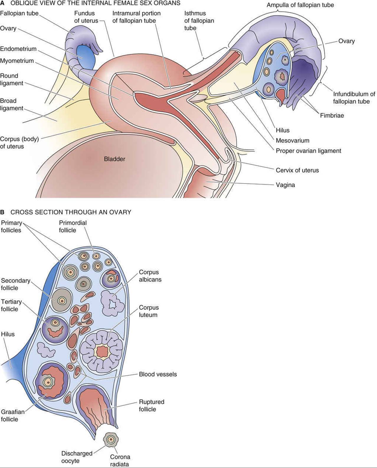
FIGURE 55-1 Anatomy of the female internal genitalia and accessory sex organs.
Female reproductive organs include the ovaries and accessory sex organs
The ovaries lie on the sides of the pelvic cavity (see Fig. 55-1A ). Covered by a layer of mesothelial cells, each ovary consists of an inner medulla and an outer cortex. The cortex of the ovary in a mature female contains developing follicles and corpora lutea in various stages of development (see Fig. 55-1B ). These elements are interspersed throughout the stroma, which includes connective tissue, interstitial cells, and blood vessels. The medulla comprises large blood vessels and other stromal elements.
The female sex accessory organs include the fallopian tubes, the uterus, the vagina, and the external genitalia. The fallopian tubes provide a pathway for the transport of ova from the ovary to the uterus. The distal end of the fallopian tube expands as the infundibulum, which ends in multiple fimbriae. The fimbriae and the rest of the fallopian tubes are lined with epithelial cells, most of which have cilia that beat toward the uterus. The activity of these cilia and the contractions of the wall of the fallopian tube, particularly around the time of ovulation, facilitate transport of the ovum. Interspersed with ciliated cells are peg cells that secrete fluid and nutrients supporting the ovum and spermatozoa as well as the zygote that may result as fertilization occurs in the fallopian tubes.
The uterus is a complex, pear-shaped, muscular organ that is suspended by a series of supporting ligaments. It is composed of a fundus, a corpus, and a narrow caudal portion called the cervix. The external surface of the uterus is covered by serosa, whereas the interior, or endometrium, of the uterus consists of complex glandular tissue and stroma. The bulk of the uterine wall consists of specialized smooth muscle, myometrium, that lies between the endometrium and the uterine serosa. The uterus is continuous with the vagina via the cervical canal. The cervix is composed of dense fibrous connective tissue and smooth-muscle cells. Glands lining the cervical canal produce a sugar-rich secretion, the viscosity of which is conditioned by estrogen and progesterone.
The human vagina is ~10 cm in length and is a single, expandable tube. The vagina is lined by stratified epithelium and is surrounded by a thin muscular layer. During development, the lower end of the vagina is covered by the membranous hymen, which is partially perforated during fetal life. In some instances, the hymen remains continuous. The external genitalia include the clitoris, the labia majora, and the labia minora, as well as the accessory secretory glands (including the glands of Bartholin), which open into the vestibule. The clitoris is an erectile organ that is homologous to the penis (see p. 1091) and mirrors the cavernous ends of the glans penis.
The breasts can also be considered as part of the female reproductive system. Breast development (thelarche) begins at puberty in response to ovarian steroid hormones. The ductal epithelium of the breast is sensitive to ovarian steroids and especially during pregnancy becomes activated to produce milk (lactation) that will sustain the newborn infant.
Reproductive function in the human female is cyclic
In some species (e.g., rabbits), female reproductive function, and specifically ovulation (the liberation of fertilizable oocytes), is triggered by mating. However, in most species, the female reproductive system functions in a cyclic manner. In some of these species with cyclic function (e.g., sheep, cattle, horses), females are receptive to males only around the time of ovulation, which maximizes the chances of fertilization and pregnancy. This receptive behavior is known as estrus, and the animals are said to have seasonal estrus cycles, whereby the ovaries are active only at a certain time of the year. Such cyclic reproductive function in females enhances reproductive efficiency by coordinating gamete production with environmental (in seasonal species) and physiological changes that attract males and prepare the reproductive tract for sperm and ovum transport, fertilization, implantation, and pregnancy. In a small subset of species (e.g., humans, baboons, apes), ovulation occurs in monthly cycles—known as menstrual cycles—that are associated with regular episodes of uterine bleeding termed menstruation.
|
LESION |
REFLEXOGENIC ERECTION |
PSYCHOGENIC ERECTION |
EFFECT ON EJACULATION |
|
Upper motor neuron |
Present |
Absent |
Significantly impaired |
|
Lower motor neuron |
Absent |
Present |
Less impaired |
Hypothalamic-Pituitary-Gonadal Axis and Control of the Menstrual Cycle
The Ovarian Cycle: Folliculogenesis, Ovulation, and Formation of the Corpus Luteum