Edward G. Moczydlowski
Physics is concerned with the fundamental nature of matter and energy, whereas the goal of medical physiology is to understand the workings of living tissue. Despite their different perspectives, physics and physiology share common historical roots in the early investigations of charge and electricity. In the late 1700s, Luigi Galvani, a professor of anatomy in Bologna, Italy, used the leg muscles of a dissected frog to assay the presence of electrical charge stored in various ingenious devices that were the predecessors of modern capacitors and batteries. He observed that frog legs vigorously contracted when electrical stimulation was applied either directly to the leg muscle or to the nerves leading to the muscle (Fig. 6-1). Such early physiological experiments contributed to the development of electromagnetic theory in physics and electrophysiological theory in biology.
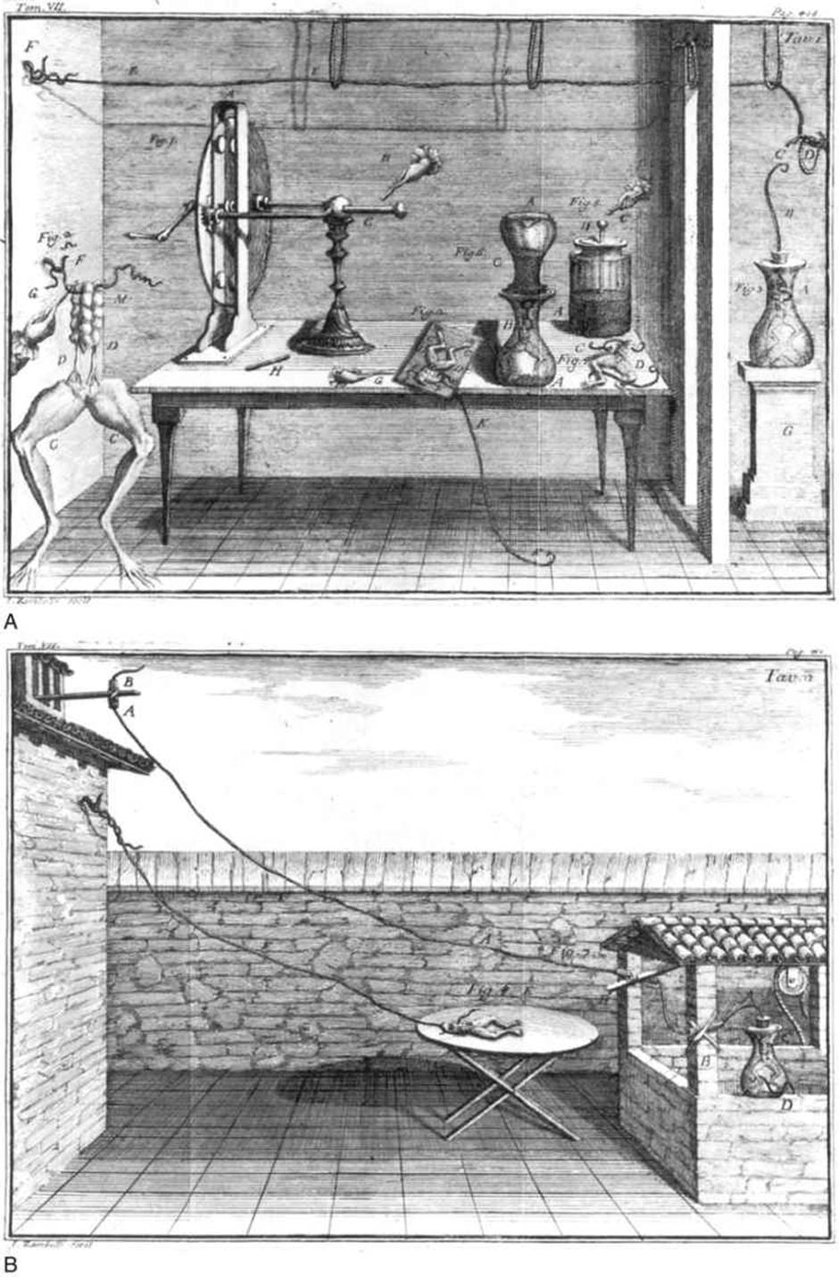
FIGURE 6-1 Early electrophysiological experiments of Galvani. For a full description, see ![]() N6-27. (From Galvani L: De viribus electricitatis in motu musculari commentarius Aloysii Galvani, Bononiae. New Haven, CT, Yale University, Harvey Cushing/John Hay Whitney Medical Library, 1791.)
N6-27. (From Galvani L: De viribus electricitatis in motu musculari commentarius Aloysii Galvani, Bononiae. New Haven, CT, Yale University, Harvey Cushing/John Hay Whitney Medical Library, 1791.)
N6-27
Early Electrophysiological Experiments of Galvani
Contributed by Ed Moczydlowski
Figure 6-1 in the text illustrates a well-known series of experiments by Luigi Galvani. A, Electrical stimulation of a dissected frog with diverse sources of electricity. On the center of the table is a board with a dissected frog that has been prepared for an experiment (Fig. Ω). A hand with a charged metal rod (G) is about to touch the sacral nerves (D), contracting the limbs (C). A metal wire (F) penetrates the spinal cord; a second metal wire (K) grounds the first wire to the floor. On the left side of the table (Fig. 1) is a large “electrical machine” with a rotating disk (A), a conductor (C), and a hand holding a metal rod (B) that is about to be charged. On the extreme left of the room (Fig. 2), a dissected frog is suspended from an iron wire that penetrates the spinal cord (F); the wire is attached to the wall by a hook. A hand with a charged metal rod (G) is touching the wire, stimulating the sacral nerves (D) and causing the legs (C) to twitch. Outside the room on the extreme right side (Fig. 3) is a frog in a glass jar (A). Emerging from the glass jar is an iron wire (B) that is attached at one end to a hook on the frog and ends in a hook (C) in the air. A silk loop (D) near this hook connects to a long conductor (F) that runs near the ceiling to a hook in the wall at the extreme left of the main room. At the far right/front of the table in the main room (Fig. 4) is a dissected frog with one conductor connected to a nerve (C) and another connected to a muscle (D). Just behind this frog (Fig. 5) is a “Leiden jar” (A) containing small lead shot used by hunters. A hand with a charged metal rod (C) is about to touch a conductor (B) emerging from the jar. To the left of the Leiden jar (Fig. 6) is an inverted jar (A) with lead shot (C). This jar sits on top of a similar jar (B) containing a suspended, dissected frog and is connected by a conductor to the lead shot in the upper jar. The legs of the frog are grounded to lead shot near the bottom of the jar. B, Electrical stimulation of the leg muscles of a dissected frog by “natural electricity” (i.e., lightning). In one experiment (Fig. 7), an iron wire (A) runs from near the roof, through several insulating glass tubes (B), to a flask (C) that contains a freshly dissected frog. A second wire (D) grounds the frog's legs to the water in the well. In a second experiment (Fig. 8), a noninsulated wire extends from an iron hook fastened to the wall and to the spinal cord of a frog (E), which is on a table coated with oil.
Reference
Galvani L. De viribus electricitatis in motu musculari commentarius Aloysii Galvani, Bononiae. Yale University, Harvey Cushing/John Hay Whitney Medical Library: New Haven, CT; 1791.
The phenomenon of “animal electricity” is central to the understanding of physiological processes. Throughout this book, we describe many basic functions of tissues and organs in terms of electrical signals mediated by cell membranes. Whereas electrical currents in a metal wire are conducted by the flow of electrons, electrical currents across cell membranes are carried by the major inorganic ions of physiological fluids: Ca2+, Na+, K+, Cl−, and ![]() . Many concepts and terms used in cellular electrophysiology are the same as those used to describe electrical circuits. At the molecular level, electrical current across cell membranes flows via three unique classes of integral membrane proteins (see pp. 17–18): ion channels, electrogenic ion transporters, and electrogenic ion pumps. The flow of ions via specific types of channels is the basis of electrical signals that underlie neuronal activity and animal behavior. Opening and closing of such channels is the fundamental process behind electrical phenomena such as the nerve impulse, the heartbeat, and sensory perception. Channel proteins are also intimately involved in hormone secretion, ionic homeostasis, osmoregulation, and regulation of muscle contractility.
. Many concepts and terms used in cellular electrophysiology are the same as those used to describe electrical circuits. At the molecular level, electrical current across cell membranes flows via three unique classes of integral membrane proteins (see pp. 17–18): ion channels, electrogenic ion transporters, and electrogenic ion pumps. The flow of ions via specific types of channels is the basis of electrical signals that underlie neuronal activity and animal behavior. Opening and closing of such channels is the fundamental process behind electrical phenomena such as the nerve impulse, the heartbeat, and sensory perception. Channel proteins are also intimately involved in hormone secretion, ionic homeostasis, osmoregulation, and regulation of muscle contractility.
This chapter begins with a review of basic principles of electricity and introduces the essentials of electrophysiology. We also discuss the molecular biology of ion channels and provide an overview of channel structure and function.
Ionic Basis of Membrane Potentials
Electrical Model of a Cell Membrane
Molecular Physiology of Ion Channels