15. Overview of Circulation
|
|
The circulatory system transports and distributes essential substances to tissues and removes metabolic byproducts. This system also participates in homeostatic mechanisms such as regulation of body temperature, maintenance of fluid balance, and adjustment of O2 and nutrient supply under various physiological states. The cardiovascular system that accomplishes these tasks is composed of a pump (the heart), a series of distributing and collecting tubes (blood vessels), and an extensive system of thin vessels (capillaries) that permit rapid exchange between the tissues and vascular channels. Blood vessels throughout the body are filled with a heterogeneous fluid (blood) that is essential for the transport processes performed by the heart and blood vessels. This chapter is a general, functional overview of the heart and blood vessels, whose functions are analyzed in much greater detail in subsequent chapters.
|
|
The heart consists of two pumps in series: one pump propels blood through the lungs for exchange of O2 and CO2 (the pulmonary circulation) and the other pump propels blood to all other tissues of the body (the systemic circulation). Flow of blood through the heart is one way (unidirectional). Unidirectional flow through the heart is achieved by the appropriate arrangement of flap valves. Although cardiac output is intermittent, continuous flow to body tissues (periphery) occurs by distention of the aorta and its branches during ventricular contraction (systole) and by elastic recoil of the walls of the large arteries with forward propulsion of the blood during ventricular relaxation (diastole).
|
|
THE CARDIOVASCULAR CIRCUIT
|
|
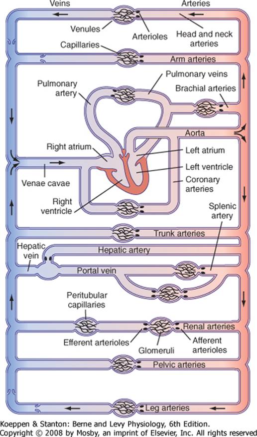
In the normal intact circulation the total volume of blood is constant, and an increase in the volume of blood in one area must be accompanied by a decrease in another. However, the distribution of blood circulating to the different regions of the body is determined by the output of the left ventricle and by the contractile state of the resistance vessels (arterioles) of these regions. The circulatory system is composed of conduits arranged in series and in parallel (Fig. 15-1). This arrangement, which is discussed in subsequent chapters, has important implications in terms of resistance, flow, and pressure in blood vessels.
|
|
Blood entering the right ventricle via the right atrium is pumped through the pulmonary arterial system at a mean pressure about one seventh that in the systemic arteries. The blood then passes through the lung capillaries, where CO2 in the blood is released and O2 is taken up. The O2-rich blood returns via the pulmonary veins to the left atrium, where it is pumped from the ventricle to the periphery, thus completing the cycle.
|
|
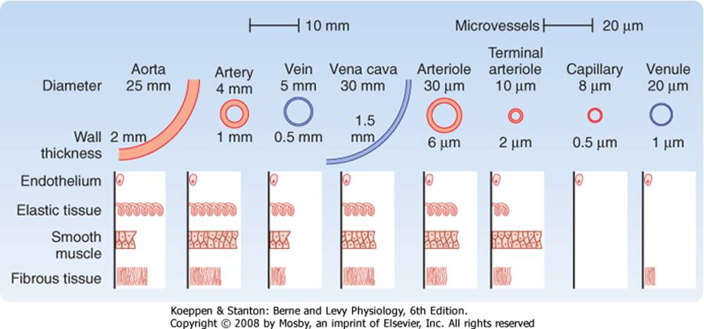
Blood moves rapidly through the aorta and its arterial branches. These branches narrow and their walls become thinner as they approach the periphery. They also change histologically. The aorta is a predominantly elastic structure, but the peripheral arteries become more muscular until at the arterioles, the muscular layer predominates (Fig. 15-2).
|
|
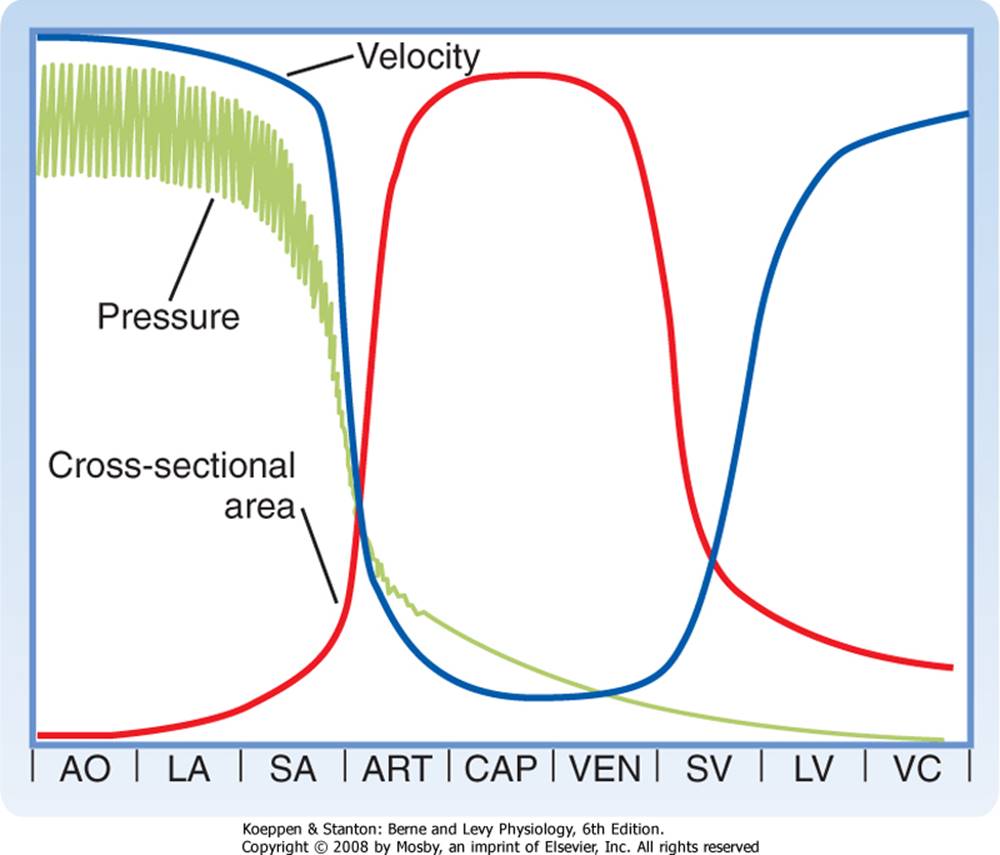
In the large arteries, frictional resistance is relatively small and pressures are only slightly less than in the aorta. The small arteries, on the other hand, offer moderate resistance to blood flow. This resistance reaches a maximal level in the arterioles, which are sometimes referred to as the stopcocks of the vascular system. Hence, the pressure drop is greatest across the terminal segment of the small arteries and the arterioles (Fig. 15-3). Adjustment in the degree of contraction of the circular muscle of these small vessels permits regulation of tissue blood flow and aids in the control of arterial blood pressure.
|
|
In addition to the reduction in pressure along the arterioles, there is a change from pulsatile to steady blood flow (Fig. 15-3). Pulsatile arterial blood flow, caused by the intermittent ejection of blood from the heart, is damped at the capillary level by a combination of two factors: distensibility of the large arteries and frictional resistance in the small arteries and arterioles.
|
|
page 289
|
|

|
|
page 290
|
|
Figure 15-1 Schematic diagram of the parallel and series arrangement of the vessels composing the circulatory system. The capillary beds are represented by thin lines connecting the arteries (on the right) with the veins (on the left). The crescent-shaped thickenings proximal to the capillary beds represent the arterioles (resistance vessels). (Redrawn from Green HD: In Glasser O [ed]: Medical Physics, vol 1. Chicago, Year Book, 1944.)
|
|
Many capillaries arise from each arteriole. The total cross-sectional area of the capillary bed is very large despite the fact that the cross-sectional area of each capillary is less than that of each arteriole. As a result, blood flow velocity becomes quite slow in the capillaries (Fig. 15-3), analogous to the decrease in velocity of flow in the wide regions of a river. Conditions in the capillaries are ideal for the exchange of diffusible substances between blood and tissue because capillaries consist of short tubes with walls that are only one cell thick and flow velocity is low.
|
|
On its return to the heart from the capillaries, blood passes through venules and then through veins of increasing size. Pressure within these vessels progressively decreases until the blood reaches the right atrium (Fig. 15-3). Near the heart, the number of veins decreases, the thickness and composition of the vein walls change (Fig. 15-2), the total cross-sectional area of the venous channels diminishes, and the velocity of blood flow increases (Fig. 15-3). Note that the velocity of blood flow and the cross-sectional area at each level of the vasculature are essentially mirror images (Fig. 15-3).
|
|
In a patient with hyperthyroidism (Graves' disease), basal metabolism is elevated and often associated with arteriolar vasodilation. This reduction in arteriolar resistance diminishes the damping effect on pulsatile arterial pressure and is manifested as pulsatile flow in the capillaries, as observed in the fingernail bed of patients with this ailment.
|
|
Figure 15-2 Internal diameter, wall thickness, and relative amounts of the principal components of the vessel walls of the various blood vessels that compose the circulatory system. Cross sections of the vessels are not drawn to scale because of the huge range from aorta and venae cavae to capillary. (Redrawn from Burton AC: Physiol Rev 34:619, 1945.)
|
|
page 290
|
|
|
|
page 291
|
|
Figure 15-3 Phasic pressure, velocity of flow, and cross-sectional area of the systemic circulation. The important features are the inverse relationship between velocity and cross-sectional area, the major pressure drop across the small arteries and arterioles, and the maximal cross-sectional area and minimal flow rate in the capillaries. AO, aorta; ART, arterioles; CAP, capillaries; LA, large arteries; LV, large veins; SA, small arteries; SV, small veins; VC, venae cavae; VEN, venules.
|
|
Data from a 20-kg dog (Table 15-1) indicate that between the aorta and the capillaries the number of vessels increases about 3 billion-fold and the total cross-sectional area increases about 500-fold. The volume of blood in the systemic vascular system is greatest in the veins and venules (67%). Only 5% of total blood volume exists in the capillaries, and 11% of total blood volume is found in the aorta, arteries, and arterioles. In contrast, blood volume in the pulmonary vascular bed is about equally divided among the arterial, capillary, and venous vessels. The cross-sectional area of the venae cavae is larger than that of the aorta. Therefore, the velocity of flow is slower in the venae cavae than in the aorta (Fig. 15-3).
|
|
Table 15-1. Vascular Dimensions in a 20-kg Dog
|
|
Vessels
|
Number
|
Total Cross-Sectional Area (cm2)
|
Total Blood Volume (%)
|
|
Systemic
|
|
Aorta
|
1
|
2.8
|
|
|
Arteries
|
40 to 110,000
|
40
|
11
|
|
Arterioles
|
2.8 × 10⁶
|
55
|
|
|
Capillaries
|
2.7 × 10⁹
|
1357
|
5
|
|
Venules
|
1 × 10⁷
|
785
|
|
|
Veins
|
110 to 660,000
|
631
|
67
|
|
Venae cavae
|
2
|
3.1
|
|
|
Pulmonary
|
|
Arteries and arterioles
|
1-1.5 × 10⁶
|
137
|
3
|
|
Capillaries
|
2.7 × 10⁹
|
1357
|
4
|
|
Venules and veins
|
2 × 10⁶ to 4
|
210
|
5
|
|
Heart
|
|
|
|
|
Atria
|
2
|
|
5
|
|
Ventricles
|
2
|
|
|
| |
|
Data from Milnor WR: Hemodynamics. Baltimore, Williams & Wilkins, 1982.
|
|
1. The circulatory system consists of a pump (the heart), a series of distributing and collecting tubes (blood vessels), and an extensive system of thin vessels (capillaries) that permit rapid exchange of substances between tissues and blood.
2. Pulsatile pressure is progressively damped by the elasticity of the arterial walls and the frictional resistance of the small arteries and arterioles such that capillary blood flow is essentially nonpulsatile. The greatest resistance to blood flow and hence the greatest pressure drop in the arterial system occurs at the level of the small arteries and arterioles.
3. The velocity of blood flow is inversely related to the cross-sectional area at any point along the vascular system.
|
|