Carrie D. Tibbles and Michael Gibbs
Pelvic fractures are a common and often life-threatening consequence of major trauma. A pelvic fracture suggests a significant force of injury, mandating a careful evaluation for multisystem involvement. Many of these patients are hemodynamically unstable, requiring aggressive volume resuscitation. These patients are at increased risk for associated injuries and significant morbidity and mortality (1). Motor vehicle collisions and automobile–pedestrian trauma are responsible for 60% to 70% of pelvic fractures. Falls from a height account for 10% to 20% of pelvic fractures, and the rest are made up of athletic and direct crush injuries (2). Early resuscitation, accurate fracture classification, stabilization of the pelvis, diagnosis of associated injuries, and coordination of key specialists are the priorities in the emergency department (ED) management of these complex injuries.
CLINICAL PRESENTATION
An understanding of both the anatomy of the pelvis and the mechanism of injury is necessary to manage patients with significant pelvic trauma. It is useful to visualize the pelvis as a ring with anterior and posterior components (Fig. 42.1). The posterior pelvic ring, which is critical for stability, is strengthened by several ligaments: the anterior and posterior sacroiliac (SI) ligaments, sacrospinous ligament, and sacrotuberous ligament (Fig. 42.2A,B). Disruption of this powerful ligamentous complex is a marker of tremendous energy transfer. Major blood vessels traversing the posterior pelvic ring are vulnerable when fractures occur here. Anteriorly, the pubic symphysis is the weakest link in the pelvic ring. Disruption of the symphysis alone does not lead to instability, provided the posterior ligamentous structures remain intact. Because the pelvis is a ring structure, if one fracture is identified, there is almost always a second disruption of the ring.

FIGURE 42.1 The pelvis.

FIGURE 42.2 Ligamentous support of the bony pelvis, (A) anterior, and (B) posterior views. The posterior pelvic ring is stabilized by several strong ligaments. Disruption in this region implies major energy transfer and the potential for significant multisystem injury.
Several different pelvic fracture classification systems have been developed. The most widely used system was developed by Young and Burgess and is based on the mechanism of injury (3). Three force vectors are described: lateral compression (LC), anteroposterior (AP) compression, and vertical shear (VS). “Complex patterns” describe injuries that are the result of more than one force vector. Each of these vectors breaks or dislocates the pelvic ring in a predictable pattern and is associated with varying degrees of mechanical instability (Table 42.1) and potential for hemorrhage. Each injury force vector has a unique effect on “pelvic volume”: that is, the potential space created after injury that may accommodate pelvic hemorrhage. Injury vectors that increase pelvic volume (e.g., AP compression, VS) are much more likely to be associated with brisk pelvic bleeding and hemodynamic instability than injury vectors that decrease pelvic volume (e.g., LC).
TABLE 42.1
Young–Burgess Classification of Pelvic Fractures

Lateral Compression
LC fractures account for almost 50% of pelvic fractures. The force is delivered to the pelvis from the side. LC fractures are common after a “T-bone” motor vehicle collision or when a pedestrian is struck from the side. The affected hemipelvis is crushed inward (Fig. 42.3). Stability is usually maintained, and therefore, LC injuries are less likely than other pelvic fractures to have associated major hemorrhage. Morbidity is secondary to coincident blunt force trauma to the head and torso, rather than the pelvic fracture itself.

FIGURE 42.3 Lateral compression injury. The causative force vector is delivered from the side, crushing the affected hemipelvis inward. Lateral compression injuries typically reduce pelvic volume. Instability is variable.
Essential radiographic features of LC fractures include horizontal pubic rami fractures. These fractures are visible 80% of time on the AP view, and this feature alone is highly suggestive of LC injury. Crush fractures of the sacrum are seen in 88% of cases. This fracture may only be visible as a disruption of the arcuate lines. The combination of a crush fracture of the sacrum and a horizontal pubic ramus fracture is diagnostic for a LC injury. Fractures of the acetabulum or hip dislocations can also be seen. SI joint diastasis in combination with any of the other findings also suggests a LC injury.
Another type of fracture that results from LC is the “stove-in” or pelvic wing fracture that was first described by Duverney in 1751 (Fig. 42.4). This fracture may produce an unstable fragment off the iliac crest, but the load-bearing stability of the pelvic ring is not jeopardized.

FIGURE 42.4 Duverney fracture. Pelvic wing or “stove-in” fracture.
Anteroposterior Compression
Nearly one-third of pelvic fractures are AP compression injuries. An anterior compressive force causes rupture of the symphyseal ligament and diastasis of the anterior pelvic ring. The posterior ligaments will tolerate up to 2.5 cm of symphyseal diastasis. Progressive widening of the symphysis beyond this point results in disruption of the anterior SI and sacrospinous ligaments. This injury is commonly referred to as an “open-book” pelvic fracture. Severe trauma will disrupt the posterior SI ligaments as well. In these circumstances, the SI joint is shown widely displaced on radiographs.
AP compression fractures are seen following head-on motor vehicle collisions or as a pedestrian is struck head-on by an oncoming vehicle. Forces may also be delivered in a posterior–anterior direction. Both iliac wings are forced outward (Fig. 42.5). AP compression injuries are unstable, and are often associated with brisk pelvic and retroperitoneal hemorrhage. Coincident injuries of the thorax and abdomen are the rule.

FIGURE 42.5 AP compression injury. The causative force vector is delivered from the front (or back), causing the pelvic ring to “open like a book.” These injuries are mechanically unstable, and associated with increases in pelvic volume.
Radiographs of anterior compression injuries will demonstrate vertically oriented pubic rami fractures, symphyseal diastasis, or both, and a variable degree of SI joint disruption. An acetabular fracture is seen in up to one half of patients.
Vertical Shear Fractures
VS injuries occur when a vertically oriented force is delivered to the pelvis via the extended femurs. These rare injuries are usually the result of a fall from a height or follow a head-on motor vehicle collision in which the occupant had the legs fully extended. VS injuries can also result from a downward force delivered to the pelvis via the spine, which may occur when a heavy object falls on the back or shoulders. Because one hemipelvis is driven up (or down) compared to the other (Fig. 42.6), these injuries produce major instability. The hemodynamic consequences are similar to those associated with AP compression injuries. Radiographs of a VS fracture demonstrate severe posterior disruption. This injury appears as either complete SI joint disruption, a vertical sacral fracture, or a medial iliac wing fracture with associated symphyseal disruption or pubic rami fractures.

FIGURE 42.6 Vertical shear injury. The causative force is transmitted up the ipsilateral femur, driving the affected hemipelvis upward. Vertical shear injuries are extremely unstable and associated with increases in pelvic volume.
The Malgaigne fracture, first described in 1859, is characterized by anterior disruption of the pubic symphysis or two to four pubic rami fractures and ipsilateral SI joint disruption. Because of involvement of the posterior arch, these injuries can be complicated by significant neurologic and vascular injury. These patients are often hypotensive, and the mortality rate approaches 20%.
Other Types of Pelvic Fractures
Straddle Injuries
A straddle injury is an unstable injury characterized by bilateral double pubic rami fractures. Classically, this fracture results from falling while straddling a solid object between the legs. Both lateral and AP compressive forces can cause straddle injuries as well. The lower genitourinary tract is particularly susceptible to injury by this mechanism. These injuries are usually closed fractures. Patients will have perineal tenderness, ecchymosis, and gross hematuria. Male patients may be unable to void. The incidence of hemorrhage and bowel injury is high.
Avulsion Fractures
These injuries are among the most trivial of pelvic fractures and are often sustained by athletes who stress the muscular or ligamentous insertions onto the bony pelvis (Fig. 42.7). Avulsions of the anterior superior iliac spine generally occur in adolescent hurdlers and long jumpers. These injuries reflect forceful contraction of the sartorius muscle or abdominal musculature transmitted to the iliac epiphysis. Less commonly, the anterior inferior iliac spine may be avulsed by intense contraction of the straight head of the rectus femoris muscle. This injury may occur in athletes playing sports that involve kicking (e.g., soccer and rugby). Avulsion fractures of the ischial tuberosity are even less common. This injury usually results from strenuous stretching of the pelvic ring, as in hurdling or pole vaulting. Patients with avulsion fractures generally present with a characteristic history and local tenderness. In isolated trauma, there are usually no associated complaints or injuries. These are stable fractures, because the integrity of the pelvic ring remains intact.

FIGURE 42.7 Fractures due to stress on muscular or ligamentous insertions onto the bony pelvis.
Sacral Fractures
Sacral fractures are defined by the orientation of the fracture line and occur in transverse upper, transverse lower, and vertical planes. Upper sacral fractures are frequently difficult to recognize both radiographically and clinically. These fractures often present only as buttock and perianal pain. These fractures may injure sacral nerve roots resulting in decreased anal sphincter tone and loss of perineal sensation. Lower sacral fractures are equally painful but are rarely complicated by neurologic injury. A careful physical examination to exclude rectal tears is important. Vertical sacral fractures occur as a result of VS injury.
Patients who fall from a standing height frequently present with pelvic pain localized to the bony prominences. Areas such as the iliac spine, ischial tuberosities, superior pubic rami, and coccyx are particularly susceptible to direct trauma. These fractures are stable injuries and are particularly common in elderly osteopenic patients. Presentation involves localized pain, swelling, and ecchymosis. There are usually no associated injuries.
Associated Injuries
Significant force is required to disrupt the pelvic ring. Consequently, associated injuries, both to adjacent viscera as well as organs distant to the pelvis, are common following major pelvic trauma. The Injury Severity Score, as a surrogate marker of organ injury, is an important predictor of mortality in patients with pelvic fractures (4).
Approximately one-fourth of pelvic fractures are complicated by injuries to the urethra or bladder (5). More than 95% of patients with an injury to the posterior urethra and 70% to 95% of patients with a bladder injury following blunt trauma have a pelvic fracture. Bladder injury is usually the result of perforation by fracture fragments and is found in patients with symphyseal diastasis and those with multiple pubic rami fractures. The posterior urethra is also prone to injury following anterior pelvic fractures because it is firmly attached to the pubis by the puboprostatic ligament.
Upper sacral fractures, VS fractures, fractures of the sciatic notch, and SI joint injuries are frequently complicated by injuries to the lumbosacral plexus. The overall incidence of neurologic injury in patients with pelvic fractures is reported to be 10% to 21%. The incidence of neurologic injury is as high as 50% in fractures with posterior disruption (4). Such patients may present with distal motor weakness (impaired dorsiflexion or plantar flexion of the great toe), numbness on the dorsal and lateral aspect of the foot, a diminished Achilles tendon reflex, or evidence of cauda equina syndrome, including perianal anesthesia and loss of sphincter tone.
Open pelvic fractures are defined as a fracture with direct communication with a vaginal, rectal, or skin wound. These fractures are invariably associated with severe injuries of the pelvic viscera. Early mortality is attributed to severe blood loss or associated chest or intracranial injuries. Historically, mortality may be as high as 30% to 50% (6). Following the initial resuscitation phase, heavy contamination of perineal wounds resulting from damage of the rectum and genitourinary tract increases the risk of delayed complications, including abscess formation, sepsis, and multiorgan failure.
ED EVALUATION
Physical Examination
During the initial resuscitation of the severely injured trauma patient, the priorities are to rapidly identify immediate life-threatening injuries and to initiate resuscitation. In the patient with a pelvic fracture, hypotension is an ominous sign, and a search for the cause should be rapidly initiated.
The objectives of the physical examination of the pelvis are to estimate the likelihood of a pelvic fracture and to identify any complications and associated injuries. The physician should inspect and palpate the front and back of the pelvis noting any abrasions, contusions, lacerations, tenderness, or crepitus. Although the identification of an unstable pelvic ring is important, overzealous rocking of the disrupted pelvis may cause additional retroperitoneal bleeding. It is preferable to apply gentle steady downward and lateral pressure on the iliac wings to detect or exclude tenderness. If tenderness is present, appropriate imaging should follow. In the obtunded blunt trauma patient with a possible pelvis injury, imaging should be performed.
Signs of urethral injury include blood at the urethral meatus, penile or scrotal hematoma, and a high-riding prostate. Patients with these findings should have a retrograde urethrogram before insertion of a Foley catheter to avoid worsening of an incomplete urethral tear. A careful vaginal examination should be performed in female patients. Perineal lacerations may have associated intravaginal mucosal tears. Bleeding is the hallmark of vaginal trauma, although it may not be immediately apparent because of muscle spasm. Vaginal injury should also be suspected if hematuria is present, or it is difficult to pass a Foley catheter.
The lumbosacral plexus is prone to injury in pelvic fractures, and a neurologic examination of the lower extremity should be performed with particular attention to the L5 and S1 nerve roots. In the patient who is awake, motor function of the L5 nerve root is assessed by dorsiflexion of the great toe against resistance. The S1 nerve root is tested by having the patient plantar flex the great toe. L5 supplies sensation to the dorsum of the foot, and S1 supplies sensation to the lateral aspect of the foot. The Achilles tendon reflex, perianal sensation, and sphincter tone should also be assessed.
Indications for Pelvic Imaging
The severely injured blunt trauma patient who is either hemodynamically unstable or obtunded or who has clinical evidence of lower abdominal or pelvic injury requires pelvic imaging. For less severely injured patients, a thorough physical examination can be used to identify the patient at low risk for pelvic fracture who can be managed safely without imaging. In a prospective trial of 2,176 alert blunt trauma patients, Gonzalez et al. (7) demonstrated that the physical examination was 93% sensitive for detecting pelvic fractures and 100% sensitive for detecting those requiring operative repair. Pelvic imaging in blunt trauma patients is recommended for patients who:
1. Are clinically unstable.
2. Have lower abdominal or pelvic tenderness.
3. Have other signs of pelvic trauma (perineal hematoma, blood at the urethral meatus, gross hematuria).
Pelvic x-Rays
A single AP radiograph will identify the large majority of pelvic fractures and can provide sufficient information to guide early management. The AP radiograph provides specific clues that will help identify the various injury patterns and fractures described in the previous section (Figs. 42.8–42.12). The film should visualize the iliac crests, each hip joint, the fifth lumbar vertebrae, and the proximal portion of each femur. Important landmarks include the pubic symphysis, superior and inferior rami, anterior superior and anterior inferior iliac spines, SI joints, sacral ala, sacral foramen, and L5 transverse processes.

FIGURE 42.8 AP plain film of a severe lateral compression injury. The left hemipelvis is crushed inward.
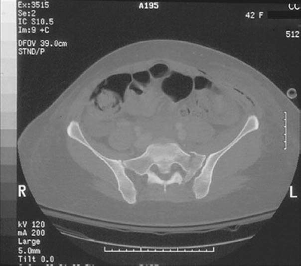
FIGURE 42.9 CT scan of a left lateral compression injury with a crush fracture of the affected sacrum.
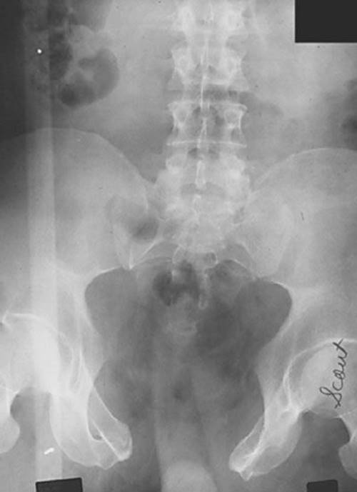
FIGURE 42.10 AP plain film of a severe AP compression injury with symphysis diastasis and left posterior SI joint disruption.

FIGURE 42.11 CT scan of an AP compression injury with major disruption of the posterior pelvic ring.

FIGURE 42.12 AP plain film of vertical shear injury with symphysis diastasis and upward migration of the left hemipelvis.
The increased use of abdominopelvic computed tomography (CT) scan in trauma patients has diminished the role of pelvic x-rays. A pelvic x-ray may be omitted in the hemodynamically stable patient who will have an abdominopelvic CT scan. An x-ray of the pelvis plays an important role in the hemodynamically unstable patient; as it may reveal the presence of an unstable pelvic fracture, suggesting a likely source of hemorrhage (8,9).
Computed Tomography
The use of CT has revolutionized the assessment of injured patients, and pelvic trauma is no exception. When compared to plain films, CT has a higher sensitivity for the detection of fracture in both adults and children. Other advantages include a more accurate assessment of pelvic instability, the ability to provide reconstructed three-dimensional images, and visualization of adjacent pelvic and abdominal viscera. Contrast extravasation seen on abdominopelvic CT is useful for the detection of arterial bleeding and accurately predicts the need for therapeutic angiography (10,11). Blackmore et al. (12) demonstrated that visualization on CT of pelvic hematoma volume greater that 500 mL was predictive of the need for angiography compared to patients with volumes less than 200 mL.
ED MANAGEMENT
Initial ED management of the severely injured patient with a pelvic fracture can be challenging and requires a multidisciplinary approach with trauma surgery, emergency medicine, orthopedics, and interventional radiology (13). Aggressive resuscitation, pelvic fracture stabilization, and determination of the most likely source of hemorrhage to guide subsequent management are the immediate priorities in the ED. In the unstable trauma patient with a pelvic injury, the critical decision is summarized as follows: Does this patient have ongoing arterial bleeding related to the pelvic fracture, therefore, warranting angiography and therapeutic embolization, or is there intra-abdominal hemorrhage where urgent laparotomy is indicated? Rapid detection of intraperitoneal hemorrhage, an understanding of fracture patterns and stability, and local protocols and available resources guide decision making.
Resuscitation
During the initial resuscitation of the patient with significant pelvic injury, the physician should anticipate hypotension and initiate early aggressive resuscitation. Adequate intravenous access and careful monitoring are crucial. A blood sample should be sent for a cross match for type-specific blood. Hemoglobin, serum lactate, and the base deficit are also used to guide resuscitation. The critically ill patient requires vigorous resuscitation with warmed crystalloid and early transfusion, as well as precautions to prevent hypothermia.
Pelvic Stabilization
Vascular injuries and substantial blood loss often complicate displaced pelvic fractures. Venous bleeding accounts for the majority of blood loss associated with pelvic fractures. Mechanically unstable injuries and those that increase pelvic volume create a potential space for ongoing hemorrhage. Pelvic stabilization can reduce pelvic volume. This practice can stabilize displaced fracture segments, which can also decrease fracture-related bleeding. Pelvic stabilization can be accomplished by several simple measures in the ED. Wrapping a sheet tightly around the pelvis will close the pelvic ring to some degree. Commercial pelvic binders are also available, and they are recommended with fractures amenable to compression, such as fractures with a widened symphysis (14).
With the advent of effective, noninvasive pelvic stabilization techniques and rapid angiography and embolization, external fixators are no longer routinely placed in the ED. External fixators may be used in the operating room in patients requiring laparotomy. When transfer to a regional trauma center is imminent, time should not be wasted waiting for an orthopedic surgeon to apply a fixator. Instead, a temporary device should be applied, and transport should proceed without delay.
Detection of Intraperitoneal Hemorrhage
During the initial assessment of the unstable patient with pelvic trauma, it is essential to quickly identify the predominant site of hemorrhage. Immediate angiography and pelvic stabilization is indicated if pelvic bleeding poses the primary life threat. An exploratory laparotomy should be performed first, however, if there is evidence of intra-abdominal hemorrhage.
Focused abdominal sonography for trauma (FAST) is routinely used in trauma centers for the rapid detection of hemoperitoneum following blunt abdominal injury. However investigators have found that the FAST examination is not sufficiently sensitive in patients with pelvic fractures to rule out intraperitoneal hemorrhage. Friese et al. reported a sensitivity of 26% for hemoperitoneum in patients with a pelvic fracture. Thirty-one of 96 patients had a false-negative study. The authors concluded that a patient with a negative FAST examination still requires a confirmatory test, such as CT or diagnostic peritoneal lavage (DPL) (15). Tayal et al. (16) also reported a lower sensitivity of the FAST examination in the presence of a pelvic fracture (80.8%) compared to patients with blunt abdominal injury without a pelvic fracture. While not sensitive in these patients, the FAST examination does have sufficient specificity in patients with unstable vital signs and a pelvic fracture to recommend laparotomy for control of intra-abdominal hemorrhage.
The following approach based on the Ultrasound (US) findings and hemodynamic status of the patient is recommended.
1. US (+), patient stable: Perform abdominal CT.
2. US (+), patient unstable: Perform DPL aspirate or laparotomy.
3. US (–), patient stable: Perform abdominal CT or close observation.
4. US (–), patient unstable: Perform DPL aspirate (17).
When using a DPL in patients with pelvic fractures, the supraumbilical technique is preferred and has higher specificity. Classically, it has been recommended that the DPL be performed using a semi-open technique, but some authors are now reporting success using a percutaneous technique. Because the lavage can be falsely positive in patients with a pelvic fracture, the DPL aspirate should be used to guide decision making. An aspirate of 10 mL of gross blood is suggestive of intra-abdominal hemorrhage (17). An algorithm for the detection of intraperitoneal hemorrhage is presented in Figure 42.13.

FIGURE 42.13 Evaluation of hemorrhage in patients with pelvic fractures.
CT imaging in the patient with pelvic trauma is straightforward, and the study of choice in the hemodynamically stable patient. Clinicians should be wary of potential delays in the unstable patient in need of immediate laparotomy or pelvic angiography.
KEY TESTING
• Plain films for hemodynamically unstable patients.
• FAST examination to assess for intraperitoneal bleeding.
• Abdominal/pelvic CT for hemodynamically stable patients.
Definitive Management
Although venous bleeding accounts for the majority of bleeding in the pelvis, injury to pelvic arteries results in brisk hemorrhage that is not controlled by pelvic stabilization alone. Arterial injury is found in up to 20% of patients with AP compression and VS injuries, but it is uncommon in LC injury (18). The most commonly injured vessels include the superior gluteal, internal pudendal, obturator, and lateral sacral arteries. In the presence of arterial injury, vascular embolization is effective in 86% to 100% of cases and improves outcome (19,20). Early mobilization of the angiography team is essential because the length of time before embolization impacts survival.
Recently, investigators have attempted to determine factors that predict which patients would benefit from early angiography and embolization. As mentioned previously, contrast extravasation and increased pelvic hemorrhage volume on CT scan are predictive of associated arterial bleeding. Blackmore et al. (21) reported that a hematocrit <30, a pulse >130, or a displaced obturator ring fracture and pubic symphyseal wide diastasis predict major hemorrhage from a pelvic fracture. Miller et al. (22) found that patients with repeated episodes of hypotension despite adequate fluid resuscitation have a high likelihood of arterial injury. Pelvic fracture patterns may be predictive of the most important source of bleeding in the unstable patient, but should not be used in isolation, but in combination with other clinical signs (18). Eastridge et al. described the injury patterns in 231 hypotensive patients with pelvic trauma. In persistently hypotensive patients with “stable” fractures (i.e., LC and AP compression injuries with minor displacement), abdominal hemorrhage was the culprit in 85% of cases. Conversely, in patients with “unstable” fracture patterns (i.e., LC and AP compression injuries with major displacement and all VS injuries), hemorrhage was predominantly from a pelvic source. Pelvic angiography was positive in 59% of cases (23). Of note, anterior acetabular fractures were recently shown to have similar rates of arterial bleeding as pelvic ring fractures (18).
Intraoperative packing of the pelvis, in an effort to tamponade venous bleeding, has become an important adjunct in the management of these patients in some trauma centers. In general, peritoneal packing is used in the unstable patient also requiring an emergent laparotomy, as a temporizing measure if angiography is not available, or as first-line procedure, with angiography following evidence of ongoing hemorrhage. Evidence suggests this technique reduces the need for transfusions and subsequent angiography (24,25).
CRITICAL INTERVENTIONS
• Aggressively resuscitate with crystalloid and early blood transfusion
• Activate a multidisciplinary team including emergency medicine, trauma surgery, interventional radiology, and orthopedics
• Rapidly detect or exclude hemoperitoneum at the bedside using US, CT scan, or diagnostic peritoneal aspirate
• Evaluate for associated visceral injuries (torso, genitourinary, and neurologic)
• Immobilize fractures that open the pelvic ring using a pelvic binder or wrapped sheet
• Initiate timely transfer to a level I trauma center
DISPOSITION
Patients with complex pelvic fractures require the resources of a level I trauma center. Rapid transport to a hospital capable of handling such injuries is essential, for these patients may deteriorate quickly. Preparation of the pelvic fracture patient for interfacility transport includes appropriate clinical stabilization, temporary pelvic stabilization, and communication with the accepting physician.
A small minority of patients with pelvic fractures can be discharged from the ED and managed as outpatients. The mechanism of injury, the age, and social supports of the patient should be factored into this decision. An example might be an isolated pubic ramus fracture in an otherwise healthy person. Consulting with an orthopedic surgeon, careful discharge instructions, and close outpatient follow-up are essential.
Common Pitfalls
• Failure to order pelvic imaging in all unconscious blunt trauma patients.
• Delays in immobilizing interventional radiology for definitive arterial embolization.
• Underresuscitation of a patient with an unstable pelvic fracture.
• Failure to diagnose associated intraperitoneal, retroperitoneal, and neurovascular injuries.
• Delaying transfer to a level 1 trauma center.
REFERENCES
1. Adams JE, Davis GG, Alexander CB, et al. Pelvic trauma in rapidly fatal motor vehicle accidents. J Orthop Trauma. 2003;17:406–410.
2. Gustavo Parreira J, Coimbra R, Rasslan S, et al. The role of associated injuries on outcome of blunt trauma patients sustaining pelvic fractures. Injury. 2000;31:677–682.
3. Langford J, Burgess A, Liporace F, et al. Pelvic fractures: Part 1. Evaluation, classification, and resuscitation. J Am Acad Orthop Surg. 2013;21(8):448–457.
4. Lunsjo K, Tadros A, Hauggaard A, et al. Associated injuries and not fracture instability predict mortality in pelvic fractures: A prospective study of 100 patients. J Trauma. 2007;62:687–691.
5. Durkin A, Sagi C, Durham R, et al. Contemporary management of pelvic fractures. Am J Surg. 2006;192:211–223.
6. Birolini D, Steinman E, Utiyama EM, et al. Open pelviperineal trauma. J Trauma. 1990;30:492–495.
7. Gonzalez RP, Fried PQ, Bukhalo M. The utility of clinical examination in screening for pelvic fractures in blunt trauma. J Am Coll Surg. 2002;194:121–125.
8. Fu C, Wang S, Hsu Y, et al. The diminishing role of pelvic x-rays in the management of patients with major torso injuries. Am J Emerg Med. 2013;32:18–23.
9. Barleben A, Jafari F, Rose J, et al. Implementation of a cost-saving algorithm for pelvic radiographs in blunt trauma patients. J Trauma. 2011;71(3):582–584.
10. Pinto A, Niola R, Tortora G, et al. Role of multidetector-row CT in assessing the source of arterial hemorrhage in patients with pelvic vascular trauma. Comparison with angiography. Radiol Med.2010;115(4):648–667.
11. Mohseni S, Talving P, Kobayashi L, et al. The diagnostic accuracy of 64-slice computed tomography in detecting clinically significant arterial bleeding after pelvic fractures. Am Surg. 2011;77(9):1176–1182.
12. Blackmore CC, Jurkovich GJ, Linnau KF, et al. Assessment of volume of hemorrhage and outcome from pelvic fracture. Arch Surg. 2003;138:504–509.
13. Khanna P, Phan H, Hardy A, et al. Multidisciplinary management of blunt pelvic trauma. Semin Intervent Radiol. 2012;29:187–191.
14. Pizanis A, Pohlemann T, Burkhardt M, et al. Emergency stabilization of the pelvic ring: Clinical comparison between three different techniques. Injury. 2013; 44(12):1760–1764.
15. Friese RS, Malekzadeh S, Shafi S, et al. Abdominal ultrasound is an unreliable modality for the detection of hemoperitoneum in patients with pelvic fracture. J Trauma. 2007;63:97–102.
16. Tayal VS, Nielsen A, Jones AE, et al. Accuracy of trauma ultrasound in major pelvic injury. J Trauma. 2006;61:1453–1457.
17. Cullinane D, Schiller H, Zielinski M, et al. Eastern Association for the Surgery of Trauma practice management guidelines for hemorrhage in pelvic fracture–update and systematic review. J Trauma.2011;71:1850–1868.
18. Magnussen R, Tressler M, Obremeskey W, et al. Predicting blood loss in isolated pelvic and acetabular high-energy trauma. J Orthop Trauma. 2007;21(9):603–607.
19. Papakostidis C, Kanakaris N, Dimitriou R, et al. The role of arterial embolization in controlling pelvic fracture haemorrhage: A systematic review of the literature. Eur J Radiol. 2012:81:897–904.
20. Tosounidis T, Giannoudis P. Pelvic fractures presenting with haemodynamic instability: Treatment options and outcomes. Surgeon. 2013:11(6):344–351.
21. Blackmore CC, Cummings P, Jurkovich GJ, et al. Predicting major hemorrhage in patients with pelvic fracture. J Trauma. 2006;61:346–352.
22. Miller PR, Moore PS, Mansell E, et al. External fixation or arteriogram in bleeding pelvic fracture: Initial therapy guided by markers of arterial hemorrhage. J Trauma. 2003;54:437–443.
23. Eastridge BJ, Starr A, Minei JP, et al. The importance of fracture pattern in guiding therapeutic decision-making in patients with hemorrhagic shock and pelvic ring disruptions. J Trauma. 2002;53:446–450.
24. Papakostidis C, Giannoudis P. Pelvic ring injuries with haemodynamic instability: Efficacy of pelvic packing, a systematic review. Injury. 2009;40:S53–S61.
25. Osborn P, Smith W, Moore E, et al. Direct retroperitoneal pelvic packing versus pelvic angiography: A comparison of two management protocols for haemodynamically unstable pelvic fractures. Injury.2009;40(1):54–60.