Like that of most masses, the differential diagnosis of testicular masses is best analyzed by the anatomic and histologic approach (Table 57). The skin may be involved by many inflammatory conditions leading to swelling, including carbuncles, cellulitis, and dermatitis of various types. Edema of the skin and subcutaneous tissue is found in cirrhosis, CHF, nephrosis, and filariasis. The tunica vaginalis is involved with hernias and hydroceles, which may be differentiated by using transillumination. The venous plexus of the scrotum and testes is involved by varicoceles and phlebitis (usually of the left venous plexus), and a varicocele may be the sign of a carcinoma of the kidney when the left spermatic vein is obstructed. Thus, one readily sees how frequently obstruction is a pathophysiologic mechanism in tumors here or elsewhere.
|
|
|
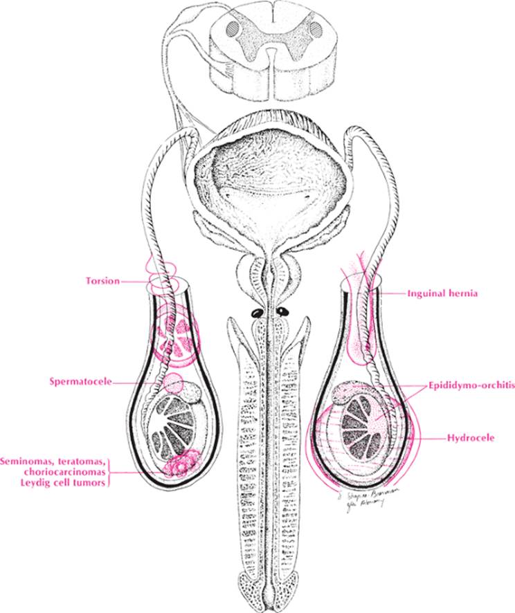
Testicular mass |
The testis is swollen in carcinomas (e.g., seminomas, choriocarcinomas, teratomas, Leydig cell tumors) and in orchitis (secondary to mumps, bacterial diseases, syphilis, or tuberculosis). The epididymis is frequently inflamed and swollen when there is orchitis and only rarely is inflamed by itself. It may also be enlarged from a spermatocele or from a vas deferens obstruction caused by prostatic disease (inflammation or neoplasm). Finally, arterial occlusion caused by torsion of the testicle may cause a testicular mass.
|
TABLE 57. Testicular Mass |
||||||||||||||||||||||||||||||||||||||||||||||||||||||||||||||||||||||||||||||||||||||||||||||||||||||||||||||||||||||||
|
||||||||||||||||||||||||||||||||||||||||||||||||||||||||||||||||||||||||||||||||||||||||||||||||||||||||||||||||||||||||
Approach to the Diagnosis
Testicular masses may be differentiated by transillumination (hydroceles and spermatoceles transilluminate, whereas hernias and tumors do not). Hernias may also be differentiated by reducing them (some will not reduce, however, if they are incarcerated), and auscultation may reveal bowel sounds. In noncommunicating hydroceles and testicular tumors, one may get above the swelling, whereas in torsion and hernias one cannot. In torsion, the tenderness is increased by elevation of the testicle, whereas in orchitis the tenderness is relieved if elevation is done for an hour or more. Serum alpha-fetoprotein beta-human chorionic gonadotropin (HCG) or lactic dehydrogenase (LDH) will be elevated in testicular tumors. Surgery may be the only way to differentiate the cause of the mass.
Other Useful Tests
1. CBC (orchitis)
2. Sedimentation rate (orchitis)
3. Urinalysis (urinary tract infection [UTI])
4. Urethral smear (infection)
5. Urine culture (UTI)
6. Urine gonadotropin (testicular tumor)
7. Prostatic fluid smear and culture (prostatitis)
8. Mumps skin test and serology (mumps orchitis)
9. Small-bowel series (hernia)
10. CT scan of the abdomen and pelvis (metastatic tumor)
11. Urology consult
12. Sonogram (torsion, hydrocele)
13. Radionuclide scan (torsion)
14. Prostate-specific antigen (PSA) (prostatic carcinoma)