D. Nathan Kim, Michael Story, and Hak Choy
For decades, radiation therapy has been a major treatment modality for locally or regionally confined cancers. The rate of treatment failure is still high, particularly for large tumors or advanced disease. Technologic improvements in radiation therapy have continuously been made that allow delivery of higher radiation doses to the tumor or lower doses to normal tissues, and in the implementation of strategies that modulate the biologic response of tumors or normal tissues to radiation. These strategies include altered fractionation scheduling including extreme hypofractionation (e.g., stereotactic body radiotherapy, stereotactic radiosurgery), combined-modality treatments using chemical or biologic agents, and, more recently, targeting molecular processes and signaling pathways that have become dysregulated in cancer cells.
The combination of chemotherapeutic drugs with radiation has perhaps had one of the strongest impacts on current cancer radiation therapy practice. This is particularly true for concurrent chemoradiation therapy, which has been shown in many recent clinical trials to be superior to radiation therapy alone in controlling locoregional disease and in improving patient survival. Combining chemotherapeutic drugs with radiation therapy has a strong biologic rationale. Such agents reduce the number of cells in tumors undergoing radiation therapy by their independent cytotoxic action and by rendering tumor cells more susceptible to killing by ionizing radiation. An additional benefit of combined treatment is that chemotherapeutic drugs, by virtue of their systemic activity, may also act on metastatic disease. Most drugs have been chosen for combination with radiation therapy based on their known clinical activity in particular disease sites. Alternatively, agents that are effective in overcoming resistance mechanisms associated with radiation therapy could be chosen. There have been clinical successes with concurrent chemoradiation therapy using traditional drugs, such as cisplatin and 5-fluorouracil (5-FU), and these studies have led to extensive research on exploring newer chemotherapeutic agents for their interactions with radiation. A number of potent chemotherapeutic agents, subsequently, have entered clinical trials or practice. These agents were selected after strong preclinical studies demonstrated that they are potent enhancers of the radiation response and thus might further improve the therapeutic outcome of chemoradiation therapy. Also, there are rapidly emerging molecular targeting strategies aimed at improving the efficacy of radiation therapy.
This chapter reviews the biologic rationale and principles fundamental to the use of chemotherapy and molecular targeted agents in conjunction with radiation treatments and discusses mechanistic interactions between drugs and radiation, the knowledge of which is essential in developing the optimal treatment strategies and designing appropriate clinical trials. It also provides a brief overview of current treatment applications and advances in the clinic. Owing to limited space, this review is far from comprehensive; additional information can be found in other reviews on this subject.1,2,3–4,5
![]() THERAPEUTIC INDEX
THERAPEUTIC INDEX
Both radiation and chemotherapeutic drugs are cytotoxic to tumor and normal tissue cells. This lack of specificity is a major limitation in their use when applied either as individual treatments or in combination. Radiation inflicts damage to tumor and normal tissues in the radiation treatment field, whereas drugs, because of their systemic action, can affect any tissue in the body. Damage is often accentuated when the two agents are combined and when they affect the same tissue. In general, both the antitumor effectiveness and the severity of normal-tissue damage produced by either radiation or drugs are increased as their dose is increased. This dose–effect relationship is sigmoidal and enables estimation of the therapeutic index (ratio), which is defined as the ratio between the doses (radiation, drug) that produce the same level (probability) of antitumor efficacy and normal-tissue damage. To be therapeutically beneficial, the therapeutic ratio must be positive (>1); that is, individual agents or their combination must be more effective against tumors than normal tissues. To define therapeutic benefit in clinical settings, many factors must be taken into account, such as whether the treatment is curative or palliative, which tissues are dose limiting (critical tissues), what degree of tissue damage is acceptable, and so forth. The balance between a given level of antitumor efficacy and acceptable normal-tissue complications gives a measure of the therapeutic ratio of a treatment.
![]() EXPLOITABLE STRATEGIES IN CHEMORADIATION THERAPY
EXPLOITABLE STRATEGIES IN CHEMORADIATION THERAPY
The goals of combining chemotherapeutic drugs with radiation therapy are to increase patient survival by improving locoregional tumor control, decrease or eliminate distant metastases, or both, while preserving organ and tissue integrity and function. Combined-modality treatment can further improve positive therapeutic outcome of individual treatments through a number of specific strategies, which Steel and Peckham6 classified into four groups: “spatial cooperation,” independent toxicity, enhancement of tumor response, and protection of normal tissues.
Spatial cooperation was the initial rationale for combining chemotherapy with radiation therapy, in which the action of radiation and chemotherapeutic drugs is directed toward different anatomic sites. Localized tumors would be the domain of radiation therapy because large doses of radiation can be given. On the other hand, chemotherapeutic drugs are likely to be more effective in eliminating disseminated micrometastases than in eradicating larger primary tumors. Thus, the cooperation between radiation and chemotherapy is achieved through the independent action of two agents. Spatial cooperation is the basis for adjuvant chemoradiation therapy, in which radiation is given first to control the primary tumor and chemotherapy is given later to cope with micrometastases. The concept of spatial cooperation is also applied in the treatment of hematologic malignancies that have spread to “sanctuary” sites, such as the brain. These sites are poorly accessible to chemotherapeutic agents, and thus they are more appropriately treated with radiation therapy.
Independent toxicity is another important strategy for increasing the therapeutic ratio of chemoradiation therapy. Normal-tissue toxicity is the main dose-limiting factor for both chemotherapy and radiation therapy. Therefore, combinations of radiation and drugs would be better tolerated if drugs were selected such that toxicities to specific cell types and tissues do not overlap with, or minimally add to, radiation-induced toxicities. This strategy requires a thorough knowledge of drug toxicity, underlying mechanisms, and drug pharmacokinetics. Another strategy in chemoradiation therapy is to exploit the ability of chemotherapeutic agents to enhance tumor radioresponse. The enhancement denotes the existence of some type of interaction between drugs and radiation at the molecular, cellular, or pathophysiologic (microenvironmental, metabolic) level, resulting in an antitumor effect greater than would be expected on the basis of additive actions. Many mechanisms may be involved in drug–radiation interactions leading to tumor radio enhancement, and some of them are elaborated on further in the text. The enhancement must be selective or preferential to tumors compared with critical normal tissues to achieve therapeutic gain. The ability of chemotherapeutic agents to enhance tumor radioresponse by counteracting determinants associated with tumor radioresistance is a major rationale for concurrent radiation therapy.
An additional strategy is to protect normal tissues so that higher doses of radiation can be delivered to the tumor. This can be achieved through technical improvements in radiation delivery or administration of chemical or biologic agents that selectively or preferentially protect normal tissues against the damage by radiation or drugs. A separate section in this chapter discusses radioprotectors in more detail.
![]() ASSESSMENT OF DRUG–RADIATION INTERACTION
ASSESSMENT OF DRUG–RADIATION INTERACTION
Any drug considered for use in combination with radiation therapy needs to undergo preclinical evaluation for its interaction with radiation both in in vitro cell culture systems and in vivo, with the aim of assessing antitumor activity and normal-tissue toxicity. The interaction between two agents is more easily defined and quantified in vitro because complete cell survival curves are readily obtained. The in vitrocell survival assay measures the ability of cells to produce colonies of a defined minimum size. Cell survival is determined after treatment with a drug or radiation alone, given at different doses, or after treatment with both agents, in which case the cells are exposed to the drug before, during, or after irradiation. Survival curves are usually plots of the surviving fraction of cells on a logarithmic scale and the dose of radiation or drugs on a linear scale.
The cell survival curve after irradiation characteristically has a “shoulder” of varying width that denotes the capacity of cells to repair radiation damage. The curves that describe survival after chemotherapeutic agents show much more variation both in absolute sensitivity to drugs and their shape than those after radiation, all depending on the drug tested. Some curves possess shoulders, some lack them, and some show resistant “tails” at higher drug doses. The tails denote the existence of cell subpopulations resistant to chemotherapeutic agents.
To assess the effect of the drug on cell radiosensitivity, the combined drug–radiation curve is commonly plotted after the cytotoxicity produced by the drug alone is excluded (“normalized”). The radiation cell survival curve is not changed if the drug does not influence cell radiosensitivity regardless of whether the drug is cytotoxic on its own. In this case, the cytotoxicity of the drug contributes only to the overall cell killing by the combined treatment (additive effect) of both agents. Chemotherapeutic agents may interact with radiation by altering cell radiosensitivity such that the combination results in a supra-additiveor subadditive effect, depending on whether the cell killing is greater or smaller than the sum of cell killings produced by individual agents. Drugs may eliminate the shoulder on the radiation survival curve, implying that drugs can inhibit cell repair from radiation damage, or they may change the slope of the exponential portion of the survival curve. A steeper slope indicates increased sensitization to radiation, whereas a shallower slope indicates protection.
Because of nonlinear dose-related characteristics in cell killing by both chemotherapeutic agents and radiation, the effects of the combined treatment are best assessed using the “isobologram,” an isoeffect plot for the dose response to the combination of two agents6 (Fig. 32.1). Dose–response curves are determined for each agent to generate the isobologram, an envelope of additivity, which denotes expected additive response over a range of doses of the agents used. If the interaction between drugs and radiation is supra-additive or synergistic (i.e., the effect is caused by lower doses of the two agents than the envelope of additivity would predict), the effect is shown at the left side of the envelope. In contrast, the effect of the subadditive or antagonistic interaction is shown to the right of the envelope: the effect required higher doses of the two agents than predicted. The width of the envelope of additivity depends on the degree of the nonlinearity in the dose response to individual agents. The envelope is wider as the degree of nonlinearity increases. In the case of a linear dose–response relationship for each agent, which is rare, the isobologram is also linear, represented by a single straight line.
In vitro testing is often followed by in vivo exploration of drug–radiation interactions, which allows assessment of the combined treatment on both tumors and normal tissues. This is essential for determination of therapeutic gain, as discussed earlier in this chapter. Syngeneic animal tumors or human tumor xenografts in nude mice are most often used for this purpose. The efficacy of the treatment is determined by the extent of tumor growth delay or the rate of tumor cure. In normal tissues, the effect of chemotherapeutic drugs on radiation response of acutely and late-responding tissues can be assessed using a variety of available assays. Some of these assays are clonogenic, such as the jejunal crypt assay, where the end point depends directly on the reproductive integrity of individual cells. More frequently, however, dose–response relationships for normal tissues are based on functional end points (such as breathing rate in lung damage and paralysis in spinal cord damage). These end points tend to reflect the minimum number of functional cells remaining in tissues or organs and not the proportion of cells retaining reproductive integrity.
FIGURE 32.1. An isobologram for two agents when their dose–response curves are nonlinear. The isobologram shows the envelope of additivity and regions of supra-additivity and subadditivity. (Modified from Steel GG, Peckham MJ. Exploitable mechanisms in combined radiotherapy-chemotherapy: the concept of additivity. Int J Radiat Oncol Biol Phys 1979;5:85–91, with permission. Copyright 1979 by Elsevier Science, Inc.)

![]() MECHANISTIC CONSIDERATIONS IN DRUG–RADIATION INTERACTIONS
MECHANISTIC CONSIDERATIONS IN DRUG–RADIATION INTERACTIONS
Increasing Initial Radiation Damage
Radiation induces many different lesions in the DNA molecule, which is the critical target for radiation damage. The lesions consist of single-strand breaks (SSBs), double-strand breaks (DSBs), base damage, DNA–DNA and DNA–protein cross-links, and so forth. DSBs and chromosome aberrations that occur in association with or as a consequence of DSBs are usually considered to be the principal damage that results in cell death.7 Any agent that makes DNA more susceptible to radiation damage may enhance cell killing. Certain drugs, such as halogenated pyrimidines, incorporate into DNA and make it more susceptible to radiation damage.8
Inhibition of Cellular Repair
Both sublethal9 and potentially lethal10,11 damage inflicted by radiation can be repaired. Although sublethal damage repair (SLDR) denotes the increase in cell survival when the radiation dose is split into two fractions of radiation separated by a time interval, potentially lethal damage repair (PLDR) designates the increase in cell survival as the result of postirradiation environmental conditions. SLDR is rapid, with a half-time of approximately 1 hour, and is complete within 4 to 6 hours after irradiation. This time between two radiation fractions allows radiation-induced DSBs in DNA to rejoin and repair. SLDR is expressed as the restitution of the shoulder on the cell survival curve for the second dose. PLDR occurs when environmental conditions prevent cells from dividing for several hours, such as keeping in vitrogrowing cells in plateau phase after irradiation. Preventing cells from division allows the completion of repair of DNA lesions that would have been lethal had DNA undergone replication within several hours after irradiation. PLDR is considered to be a major determinant responsible for radioresistance in some tumor types, such as melanomas. The repair can be achieved through restoration of damaged molecules by reducing species that donate electrons to oxidized substrates or through involvement of enzymes mediating homologous and nonhomologous recombination repair of DNA DSBs, base excision repair of base damage, and nucleotide excision repair of DNA–protein cross-links.
Many chemotherapeutic agents used in chemoradiation therapy interact with cellular repair mechanisms and inhibit repair, and hence may enhance cell or tissue response to radiation. The aforementioned halogenated pyrimidines enhance cell radiosensitivity not only through increasing initial radiation damage but also by inhibiting cellular repair.8,12 Nucleoside analogs, such as gemcitabine, are a class of chemotherapeutic agents potent in inhibiting the repair of radiation-induced DNA and chromosome damage.13,14 They have been shown strongly to enhance tumor radioresponse in preclinical studies and have been, and are continuing to be, extensively investigated for such activity in patients with cancer.15,16
Cell Cycle Redistribution
Both chemotherapeutic agents and radiation are more effective against proliferating than nonproliferating cells. Their cytotoxic action further depends on the position of cells in the cell cycle. Cell cycle dependency in response to radiation was first described almost 40 years ago.17 Terasima and Tolmach.17 reported that the sensitivity of the cell response to radiation varied widely depending on which phase of the cell cycle the cells were in at the time of irradiation, and that cells in the G2 and M cell cycle phases were approximately three times more sensitive than cells in the S phase. The exact reason for this variability is still unknown.
The influence of the cell cycle on cell response to cytotoxic agents can be therapeutically exploited in chemoradiation therapy using cell cycle redistribution strategies. For example, some chemotherapeutic drugs, such as taxanes, can block transition of cells through mitosis, with the result that cells accumulate in the radiosensitive G2 and M phases of the cell cycle. Radiation delivered at the time of significant accumulation of cells in both the G2 and M phases results in enhanced radioresponse of cells in vitro18,19 and of tumors in vivo.20,21 However, this cell cycle mechanism of taxane-induced enhancement of tumor radioresponse is dominant only in tumors that are resistant to paclitaxel or docetaxel as a single treatment. Although tumor growth in taxane-resistant tumors is not substantially affected by the drug, tumors do exhibit significant transient accumulation of cells in mitosis 6 to 12 hours after the treatment.21 Taxanes also enhance the radioresponse of tumors that respond by significant tumor growth delay to taxanes given as a single treatment modality, but a major mechanism for radio enhancement in such tumors is reoxygenation of radioresistant hypoxic cells, as discussed later.20
Elimination of the radioresistant S-phase cells by the chemotherapeutic agents may be another cell cycle redistribution strategy in chemoradiation therapy. Nucleoside analogs, such as fludarabine or gemcitabine, are good examples of the agents that become incorporated into S-phase cells and eliminate them by inducing apoptosis.13,15 In addition to purging S-phase cells, the analogs induce the surviving cells to undergo parasynchronous movement to accumulate in the G2 and M phases of the cell cycle between 1 and 2 days after drug administration, a time when the highest enhancement of tumor radioresponse was observed.15 Tumors with a high cell growth fraction are likely to respond better to the cell cycle redistribution strategy in chemoradiation therapy than tumors with a low cell growth fraction.
Counteracting Hypoxia-Associated Tumor Radioresistance
Solid malignant tumors usually are characterized by defective vascularization, both in the number of blood vessels and vessel function. Because of this, blood supply to tumor cells is inadequate, cells lack oxygen and nutrients, and multiple tumor microregions become hypoxic, acidic, and eventually necrotic. Hypoxia occurs at distances from blood vessels larger than 100 to 150 μm. The hypoxic cell content in tumors varies widely and can be more than 50%. The presence of hypoxia makes tumors more aggressive (hypoxia is conducive to the emergence of more virulent tumor cell variants and stimulates metastatic spread22,23) and more resistant to radiation as well as most chemotherapeutic agents. Hypoxic cells are 2.5 to 3 times more resistant to radiation than well-oxygenated cells. The fact that hypoxia may be a cure-limiting factor in radiation therapy, at least in some clinical situations, is suggested by the findings that reduced hemoglobin levels24 and low tumor pO225,26 are associated with higher treatment failure rates. Also, there are reports showing that local tumor control by radiation therapy can be improved by the use of hypoxic cell radiosensitizers27 or hyperbaric oxygen.28,29 With respect to the effects of chemotherapy, hypoxic regions are less accessible to chemotherapeutic drugs; in addition, hypoxic tumor cells are either nonproliferating or they proliferate poorly, and as such do not respond well to drugs.
Combining chemotherapeutic agents with radiation therapy can reduce or eliminate hypoxia or its negative influence on tumor radioresponse. Most chemotherapeutic drugs preferentially kill proliferating cells, primarily found in well-oxygenated regions of the tumor. Because these regions are located close to blood vessels, they are easily accessible to chemotherapeutic agents. Destruction of tumor cells in these areas leads to an increased oxygen supply to hypoxic regions, and hence reoxygenates hypoxic tumor cells. Massive loss of cells after chemotherapy lowers the interstitial pressure, which then allows the reopening of previously closed capillaries and the re-establishment of blood supply. It also causes tumor shrinkage so that previously hypoxic areas are closer to capillaries and thus accessible to oxygen. Finally, by eliminating oxygenated cells, more oxygen becomes available to cells that survived chemotherapy. It was recently shown that tumor reoxygenation is a major mechanism underlying the enhancement of tumor radioresponse induced by taxanes in tumors sensitive to these drugs.20
Another approach to counteract the negative impact of hypoxia is selective killing of hypoxic cells through bioreductive drugs, such as tirapazamine,22 which undergo reductive activation in a hypoxic milieu, rendering them cytotoxic. A related possibility is to exploit the acidic state (low pH) of tumors, which develops as a result of hypoxia-driven anaerobic metabolism that produces lactic acid,30 through the use of drugs that selectively accumulate in acidic environments or become activated by a low pH.31
The use of agents that selectively radiosensitize hypoxic cells to reduce their negative impact has been considered and tested in clinical trials for some time. These drugs increase radiation damage by mimicking the effect of oxygen. Many clinical trials have tested these drugs, particularly misonidazole, in combination with radiation therapy, but few of them have shown an improved treatment outcome. The exception is nimorazole, which also does not elicit the neurotoxicity commonly associated with hypoxic cell sensitizers that prevented the delivery of clinically effective doses of these agents.32
Inhibition of Tumor Cell Repopulation
The constant balance between cell production and cell loss maintains the integrity of normal tissues. When this balance is perturbed by cytotoxic action of chemotherapeutic drugs or radiation, the integrity of tissues is re-established by an increased rate of cell production. The cell loss after each fraction of radiation during radiation therapy induces compensatory cell regeneration (repopulation), the extent of which determines tissue tolerance to radiation therapy. In contrast to normal tissues, malignant tumors are characterized by an imbalance between cell production and cell loss in favor of cell production. As with normal tissues, tumors also respond to radiation- or drug-induced cell loss with a compensatory regenerative response. Preclinical studies provided ample evidence demonstrating that the rate of cell proliferation in tumors treated by radiation or chemotherapeutic drugs is higher than that in untreated tumors.33–35 This increased rate of treatment-induced cell proliferation is commonly termed accelerated repopulation. Accelerated repopulation of tumor clonogens has been shown to occur during clinical radiation therapy as well. Withers et al.36 showed that the total dose of radiation needed to control 50% of head and neck carcinomas progressively increased with time whenever radiation therapy treatment was prolonged beyond 1 month. This increase in radiation dose required to achieve tumor control was greater than what would be anticipated based on the pretreatment tumor volume doubling time of approximately 60 days for head and neck tumors. The increase was attributed to accelerated repopulation, and it was estimated to average approximately 0.6 Gy/day,36 but may be as high as 1 Gy/day.37
Although accelerated cell proliferation is beneficial for normal tissues because it spares them from radiation damage, it has an adverse impact on tumor control by radiation therapy or chemotherapy. Chemotherapeutic drugs, because of their cytotoxic or cytostatic activity, can reduce the rate of proliferation when given concurrently with radiation therapy, and hence increase the effectiveness of the treatment. Caution must be taken to select drugs that preferentially affect rapidly proliferating cells and preferentially localize in malignant tumors. However, the main limitation of concurrent chemoradiation therapy is the enhanced toxicity of rapidly dividing normal tissues because most available chemotherapeutic agents show poor tumor selectivity. Moreover, accelerated repopulation induced by chemotherapeutic drugs may have a negative influence on the outcome of tumor response to radiation when drugs are used in induction or neoadjuvant chemotherapy protocols. In this strategy, chemotherapy precedes radiation therapy. Treatment outcomes after induction chemotherapy followed by radiation therapy have not been overly encouraging in terms of both local tumor control and patient survival, even if a large proportion of tumors initially responded with total or partial clinical regression by the time of radiation therapy implementation. Some experimental evidence suggests that the drug-induced accelerated cell repopulation can actually make the tumor more difficult to control with radiation.33,34
Other Potential Interactions
Molecular Signaling Pathways That May Be Responsible for Radioresistance. Significant strides have been made in elucidating molecular pathways that may be involved in resistance to cytotoxic therapy including radiation treatments. These molecular determinants are being scrutinized in preclinical and clinical settings as potential targets for cancer therapy, of which some agents are being studied for the potential for enhancing radiation effects.38 For example, the efficacy of combining the epidermal growth factor receptor (EGFR) inhibitor cetuximab with radiation therapy has been demonstrated in randomized clinical studies in head and neck cancer patients.39 Molecular targeting has become an immensely important topic, and effective integration of these strategies with radiation therapy to foster improved efficacy of therapy is an active area of investigation. This topic is further discussed in significant detail in the section to follow.
Targeting the Tumor Microenvironment. Tumor microenvironment has been postulated as being a potential therapeutic target for cancer therapy. Tumor microenvironment is a complex system of many cell types including endothelial cells, smooth muscle cells, fibroblasts, and cells involved with the immune system (lymphocytes, macrophages, etc.).40 Among these different cells that make up the different components of the tumor microenvironment, the most studied preclinically and clinically are the cells that make up the tumor microvasculature. Preclinical studies have suggested the potential role for radiosensitization of the tumor microvasculature using compounds directed at targeting angiogenesis.41 Clinical studies have been performed with efforts to combine antiangiogenic agents with radiation therapy, with mixed results.41 Studies are ongoing, as many antiangiogenic agents are approved for cancer therapy, and are detailed in the section on antiangiogenesis agents.
Cancer Stem Cells. Cancer stem cells are defined as being cells within a tumor that possess the capacity to self-renew and generate the heterogeneous lineages of cancer cells that make up the tumor.42 Cancer stem cells as a source of radiation resistance for solid tumors is an area of active investigation. Potential for the need to eradicate cancer stem cells for effective cure by radiotherapy has been postulated, and while radioresistance of cancer stem cells has been noted in in vitro studies,43 this notion has been challenged as some have demonstrated radiosensitivity of cancer stem cells.44 But, if in fact cancer stem cells are radioresistant, one possible strategy may be to identify molecular pathways that could influence cancer stem cell radiosensitivity and investigate the use of such agents in combination with radiotherapy at different stages during the course of therapy.45
TABLE 32.1 ADVANTAGES AND DISADVANTAGES OF DIFFERENT CHEMORADIATION SEQUENCING STRATEGIES
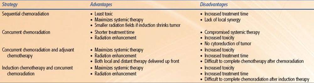
TABLE 32.2 MECHANISMS OF CHEMOTHERAPY-INDUCED RADIATION SENSITIZATION

![]() TIMING OF DRUG ADMINISTRATION IN RELATION TO RADIATION THERAPY
TIMING OF DRUG ADMINISTRATION IN RELATION TO RADIATION THERAPY
Most clinical chemoradiation therapy regimens evolved empirically: increasingly, information from preclinical studies is being considered in planning the optimal timing of drug administration in relation to radiation therapy. Depending on the principal aim of the therapy, drugs are administered before (induction or neoadjuvant chemotherapy), during (concurrent or concomitant chemotherapy), or after (adjuvant chemotherapy) the course of radiation therapy. The advantages and disadvantages of each approach are summarized in Table 32.1.
In regard to the primary tumor, induction chemotherapy may reduce the number of clonogenic cells and cause the reoxygenation of the surviving hypoxic cells, both of which render tumors more controllable by radiation. In addition, chemotherapy-induced tumor shrinkage may allow the use of smaller radiation fields, in which case less normal tissue is exposed and damaged by radiation. This treatment approach is often used in the therapy of solid tumors in children and of lymphomas. Induction chemotherapy precedes radiation therapy for a few weeks to a few months, which improves tolerability of the combined treatment.
Induction chemotherapy has resulted in therapeutic improvement in a number of clinical trials compared with radiation therapy, but in general the therapeutic benefits are below expectations. A number of factors could account for this, including accelerated proliferation of tumor cell clonogens and selection or induction of drug-resistant cells that are cross-resistant to radiation. The preclinical findings provide solid evidence for the existence of accelerated repopulation in tumors treated with chemotherapeutic agents. On the other hand, although development of drug resistance is a significant problem in chemotherapy, the evidence that cells that acquire drug resistance are also resistant to radiation is not convincing.
When chemotherapy is given during a course of radiation therapy, it is referred to as concurrent chemotherapy. This form of treatment is intended to cope with both disseminated lesions and the primary tumor, but it takes advantage of drug–radiation interactions to maximize tumor radioresponse. The drug scheduling in relation to individual radiation fractions is highly important, and the selection of optimal timing of drug administration must be based on mechanisms of tumor radio enhancement by a given drug, the drug’s normal tissue toxicity, and the conditions under which the highest enhancement is achieved. The data from preclinical studies can greatly contribute to the selection of the most optimal schedules. For example, it has been demonstrated that murine tumors sensitive to taxanes show enhanced radioresponse, but the best effect is achieved if drug treatment precedes radiation by 1 to 3 days.20 A major mechanism for tumor radio enhancement was reoxygenation of hypoxic cells. Based on this preclinical information, one would anticipate that in clinical protocols such tumors would best respond to a bolus of a taxane given once or twice weekly during radiation therapy. In contrast, tumors resistant to taxanes on their own would call for daily administration of a taxane because they show accumulation of radiosensitive G2- and M-phase cells 6 to 12 hours after drug administration. If the objective is to counteract rapid repopulation of tumor cell clonogens induced by radiation, then administration of cell cycle–specific chemotherapeutic agents during the second half of radiation therapy, when accelerated repopulation is more expressed, might be more effective. At present, the enhancement in normal-tissue complications remains the major limitation of concurrently combining chemotherapy with radiation therapy. Nevertheless, as is made evident later in the text, concurrent chemoradiation therapy has provided better clinical results both in terms of local tumor control and patient survival than have other modes of chemoradiation therapy combinations.46,47
Adjuvant chemotherapy designates a treatment modality in which chemotherapeutic drugs are given some time after completion of radiation therapy. The primary objective is to eradicate disseminated disease; however, the control of the primary tumor may also be improved by the ability of drugs to deal with tumor cells that survived radiation.
![]() INTERACTION OF SPECIFIC CHEMOTHERAPIES AND RADIATION IN THE TREATMENT OF CANCER
INTERACTION OF SPECIFIC CHEMOTHERAPIES AND RADIATION IN THE TREATMENT OF CANCER
This section provides an overview of the evidence that exists for combining particular chemotherapies with radiation. In many cases, the level of support that exists for combined therapy mirrors the age of the drug. However, as would be expected, newer drugs have generally been subject to more rigorous preclinical assessment of their efficacy before their introduction into the clinical setting (Table 32.2).
Platinum-Based Drugs
This group of compounds, distinguished from most others by its metallic element base, has come to be recognized as one of the most potent chemotherapies available to date. Cisplatin (cis-diamminedichloroplatinum II), which is the prototype drug, has been acknowledged to be a potent radiosensitizer for many years and has a significant role in clinical practice to date. Preclinical work done using murine models by Rosenberg et al.64 in the late 1960s showed that cisplatin is an effective antitumor chemotherapy. Subsequent efforts have shown that its primary mechanism of inhibition of tumor growth appears to involve the inhibition of DNA synthesis.65,66 Another secondary mechanism includes the inhibition of transcription elongation by DNA interstrand cross-links.67
Work on nonmammalian systems first demonstrated the radiosensitizing abilities of platinum-based compounds.68–70 This was confirmed in several mammalian systems as well.48,71,72 This makes inherent sense because these platinum compounds have a high electron affinity and react preferentially with hydrated electrons. The exact mechanism for the increased cell death seen with combinations of ionizing radiation and platinum drugs is not known for certain; however, the evidence would seem to point to the inhibition of PLDR49 and to the radiosensitization of hypoxic tumor cells.73 Cisplatin free radical–mediated sensitization may involve the ability to scavenge free electrons formed by the interaction between radiation and DNA. The reduction of the platinum moiety may serve to stabilize DNA damage that would otherwise be repairable.74 Greater than additive effects of cisplatin and radiation are seen in tumor models most reliably when the drug is administered with fractionated radiation,74 explained by its inhibition of SLDR.
Carboplatin, a second-generation platinum compound with a different toxicity profile, has also been studied as a radiosensitizer.50,51 Its potential efficacy as a radiosensitizer has allowed for its incorporation into regimens used in several randomized trials. Interest exists in the combination of radiation with other platinum analogs, including oxaliplatin75 as well as orally administered compounds like satraplatin.76–78
Oxaliplatin, although sharing a similar mode of action as other platinum compounds, has been shown to have activity in cisplatin-resistant systems in vitro.4 One potential explanation for this is that oxaliplatin is not affected by loss of mismatch repair, which leads to cisplatin resistance in vitro,79 and there are those who postulate that adducts formed by oxaliplatin are less well recognized by DNA repair systems.80 Whether oxaliplatin is truly active against cisplatin-resistant cancer clinically is an area of active investigation,81 and after encouraging findings in phase I to II rectal cancer studies, it entered into phase III studies, some of which have closed to accrual.80
Antimicrotubules
Taxanes
The radiosensitizing properties of plant-derived chemotherapeutic agents, the taxanes, have been studied extensively in both preclinical models and in clinical trials. Paclitaxel (Taxol) and docetaxel (Taxotere) act as mitotic spindle inhibitors through their promotion of microtubule assembly and inhibition of disaggregation.82 Both taxanes bind to the N-terminal 31–amino-acid sequence of the β-tubulin subunit of cellular tubulin polymers, stabilizing the polymers by shifting the dynamic equilibrium that exists between tubulin dimers and microtubules in favor of the polymerized state.83,84 Although there is preclinical evidence that docetaxel has both a higher affinity for the tubulin-binding site and greater in vitro cytotoxicity than paclitaxel, this has not necessarily translated into greater clinical efficacy because the toxicity profiles of the two drugs also differ.85,86
The administration of a taxane leads to cellular arrest in the G2/M phase of the cell cycle, which is the precise point associated with increased sensitivity to the lethal effects of ionizing radiation.87 Early laboratory studies with a human lung cancer cell line19 and human astrocytoma cells18 bore out the prospect of significant radiosensitization, with relative enhancement ratios in the 1.48 to 1.8 range when paclitaxel was administered before irradiation.
The exact conditions used in various studies appear to determine the strength of the interaction between radiation and paclitaxel because subadditive effects have been seen in addition to the more widely reported additive and supra-additive effects.21 In general, enhancement of radiation effects is seen when proliferating cells have been incubated with moderate concentrations of paclitaxel for 24 hours before irradiation. Conditions leading to a less-than-optimal response include paclitaxel-mediated G1 arrest, wherein a more resistant cell subpopulation counteracts the effects of a G2/M block; paclitaxel-induced cell cycle effects such as the G2/M block in cells destined to die before irradiation; and incubation conditions insufficient to exert cellular effects. The fact that nonproliferating cells are also sensitized to the effects of radiation by the use of paclitaxel suggests that mechanisms other than the cell cycle arrest in the G2/M phase underlie paclitaxel’s sensitizing abilities.
Paclitaxel also acts to induce programmed cell death; work from the M.D. Anderson Cancer Center21 has examined the relationship between mitotic arrest, apoptosis, and the antineoplastic activity of paclitaxel in 16 murine tumors. Single-dose paclitaxel (40 mg/kg) induced mitotic arrest to varying degrees in all tumors; however, apoptosis was induced in only 50% of tumors. This study also revealed that pretreatment levels of apoptosis correlated with both paclitaxel-induced apoptosis and tumor growth delay. Therefore, both the pretreatment apoptotic rate and paclitaxel-induced apoptotic rate could potentially act as predictors of the response to paclitaxel.
Milas et al.21 summarized observations that showed that (a) there was massive loss of tumor cells though the apoptotic pathway was restricted to the perivascular region, and (b) radio enhancement occurring during this period of cell loss became even more impressive when apoptotic cells were removed from the tumor. Experiments in which tumor xenografts were treated with paclitaxel and exposed to radiation under hypoxic or air-ambient conditions were pursued.20 It was found that the creation of hypoxic conditions greatly reduced the efficacy of paclitaxel in its enhancement of radioresponse. It appears that a combination of cell cycle effects, drug-induced reoxygenation, and drug-induced apoptosis underlies paclitaxel’s radiosensitizing abilities.
Docetaxel has also been found to be a respectable radiosensitizer in both in vitro52 and in vivo models.53 Radiation response in the presence of docetaxel was examined in three different cell lines that have widely different responses to radiation alone.88 Their findings suggest that the p53 status of tumor cells may have a profound effect on the radiosensitizing effects of a taxane. Other novel taxanes and analogs that continue to attract the interest of investigators for their potential to enhance radiation effects include Abraxane, paclitaxel poliglumex, larotaxel, cabazitaxel,89,90 and orally available taxanes.91
Epothilones
Epothilones are a novel class of antimicrotubule agents, originally derived from the myxobacterium Sorangium cellulosum. These agents bind to the site near the taxane binding site, and mechanism of action is similar to taxanes89,92,93; however, their chemical structures are unrelated, and their binding is specific and independent of the taxanes. Several epothilones are undergoing investigation in clinical trials: patupilone, ixabepilone, BMS-310705, ZK-EPO, and epothilone D.94 Epothilones enhance microtubule stability, formation of abnormal mitotic spindles inducing G2 and M arrest, and apoptosis. Interestingly, patupilone has been shown to cross the blood–brain barrier, and studies with concurrent radiation therapy for central nervous system (CNS) malignancies have been performed at the phase I level.95 The potential for patupilone to work as a radiosensitizer for multidrug-resistant cancer cells in vitrohas been reported,96 as well as for medulloblastoma cells,97 lung cancer, and prostate cancer cells.98,99
Antimetabolites
5-Fluorouracil (5-FU)
The radiosensitizing properties of 5-FU have been known for years.100 Several mechanisms have been proposed for the cytotoxicity of this drug:
a. Its incorporation into RNA, which leads to a disruption of RNA function;
b. Inhibition of thymidylate synthetase function and subsequently of DNA synthesis; and
c. Direct incorporation of the drug into DNA.
It is believed that a combination of these effects underlies its radiosensitizing properties.101 Optimization of its schedule of delivery is crucial to obtaining an effect with this combination, and it is accepted that cytotoxic doses of 5-FU are needed to obtain a radiosensitizing effect. In general, it is thought that a continuous infusion of the drug is needed to obtain the desired drug levels after irradiation.102 Long-standing clinical experience with this drug bears out much of the laboratory studies of its effectiveness as a radiation sensitizer.
Capecitabine
Capecitabine is an oral prodrug of 5-FU. It is converted to its cytotoxic form in three enzymatic steps, the last of which is mediated by thymidine phosphorylase. One of the potential advantages of this mechanism for increasing tumor cytotoxicity is that thymidine phosphorylase is overexpressed in tumor tissues. Interestingly, radiation has been shown to stimulate expression of thymidine phosphorylase, which provides a further rationale for considering combined therapy with radiation treatments.103 Several phase I/II studies of radiation therapy and capecitabine primarily in rectal cancer have been completed,104,105 and a large phase III study from the National Surgical Adjuvant Breast and Bowel Project (NSABP) is under way.105
Gemcitabine
Gemcitabine is another nucleoside analog that acts as a very potent radiosensitizer. The biologic action of gemcitabine is due almost completely to its effects on DNA metabolism. Early studies of this drug in leukemic cell lines found that notable decreases in cellular deoxynucleotide triphosphates occurred with the use of the drug.106
Direct incorporation of the drug into DNA and drug-induced apoptosis are also thought to underlie its cytotoxicity.106 The metabolism of gemcitabine in the cell is complex, and it is able to potentiate its cytotoxicity as a sole therapy.106 Depending on the conditions, relative enhancement ratios in the range of 1.1 to 2.5 have been reported.
Gemcitabine is S-phase specific and as such should be selectively toxic to proliferating cells,107 decreasing the amount of proliferation that can occur during fractionated radiation therapy. In addition, cell cycle redistribution induced by these agents may improve cell kill by allowing more cells to be treated in the more sensitive parts of the cell cycle.108 As DNA synthesis inhibitors, these drugs may act to inhibit the repair of radiation-induced DNA damage.13 Finally, as DNA chain terminators, they may serve to trigger the apoptotic response.14
There is strong preclinical evidence to suggest that the radiosensitizing abilities of gemcitabine are intimately linked to cellular deoxyadenosine triphosphate levels.14 Interesting results from Latz et al.60 show quite clearly that cells that are pretreated with gemcitabine no longer show a progressive increase in radioresistance as they progress toward DNA replication, and sensitization, therefore, appears to be greatest in the S phase.
Milas et al.15 found the largest enhancement of growth delay when gemcitabine was delivered 24 to 60 hours before irradiation in a murine sarcoma tumor model. The use of gemcitabine with radiation also decreased the risk for development of lung metastases in those mice that attained durable local control (73% in the radiation-alone group vs. 40% in the combined-modality group), which was confirmed in a second study with a larger number of mice.61This preclinical work is supportive of the principles of combined-modality therapy in that better local control translated into decreased systemic spread of tumor cells. The same authors also report a dose-dependent increase in the apoptotic rate after the administration of gemcitabine,15 which they believe correlates with the elimination of the more radioresistant S-phase population of cells and a redistribution of the remaining cells into more radiosensitive parts of the cell cycle. They also report that reoxygenation of the resistant hypoxic fraction of tumor cells is a mechanism for the radiosensitizing action of gemcitabine.61
In summary, the preclinical evidence suggests that gemcitabine acts through several mechanisms (nucleotide pool perturbation, lowering of the apoptotic threshold, cell cycle redistribution, and tumor cell reoxygenation) to enhance the effect of ionizing radiation on tumors. Clinical experience has shown that this drug is indeed a potent sensitizer with the potential for significant toxicity as well as improvement in tumor control when combined with radiation.109,110,111–115
Pemetrexed
Pemetrexed is a novel multitargeted agent, exerting its effect via simultaneous inhibition of multiple folate-requiring enzymes, including thymidylate synthase, dihydrofolate reductase, and glycinamide ribonucleotide formyl-transferase.116 Pemetrexed has shown synergistic activity with radiation treatments, likely due to interference with DNA synthesis.117 There are indications that radiosensitization by pemetrexed is not cell cycle phase specific118 in vitro. Others have suggested that a combination of radiation therapy and pemetrexed results in supra-additive effects, which may be in part due to apoptosis induction.119 Phase I/II studies in non–small cell lung cancer (NSCLC) and esophageal cancer combining pemetrexed-based chemotherapy with radiation therapy have been completed demonstrating tolerability and encouraging results.120,121,122,123–126
Topoisomerase I Inhibitors
Camptothecin is a plant alkaloid obtained from the Camptotheca acuminata tree. Its initial clinical evaluation in the 1960s and 1970s was abandoned because of severe and unpredictable hemorrhagic cystitis.127,128 Camptothecin and its derivatives (e.g., irinotecan, topotecan, 9-aminocamptothecin, SN-38) target DNA topoisomerase I.129–130,131 This enzyme relaxes both positively and negatively supercoiled DNA and allows for diverse essential cellular processes, including replication and transcription, to proceed. In the presence of camptothecin, a camptothecin–topoisomerase I–DNA complex becomes stabilized with the 5′-phosphoryl terminus of the enzyme-catalyzed DNA SSB bound covalently to a tyrosine residue of topoisomerase I. These stabilized cleavable complexes interact with the advancing replication fork during the S phase or during unscheduled DNA replication after genomic stress and cause the conversion of SSBs into irreversible DNA DSBs, resulting in cell death.130
Several investigators have reported that camptothecin enhances the cytotoxic effect of radiation in vitro and in vivo.54–55,56 Chen et al.55 showed that cells exposed to 20(S)-10,11 methylenedioxycamptothecin before or during radiation had sensitization ratios of 1.6, whereas those treated with the drug after radiation had substantially less enhancement of radiation-induced DNA damage. There are several hypotheses regarding the mechanism of interaction between radiation and irinotecan: (a) inhibition of topoisomerase I by irinotecan leads to inhibition of repair of radiation-induced DNA strand breaks; (b) irinotecan causes a redistribution of the cells into the more radiosensitive G2phase of the cell cycle; (c) topoisomerase I–DNA adducts are trapped by irinotecan at the sites of radiation-induced SSBs, leading to their conversion into DSBs.57 While there is currently insufficient evidence to identify the underlying mechanism with certainty, the primary mechanism involved with radiosensitization may depend on which camptothecin derivative is being used.
Data from in vivo experiments demonstrate that combination 9-aminocamptothecin and irradiation is more effective when fractionated, compared with single doses.132 There is also evidence for circadian-dependent cytotoxicity and radiation sensitization when camptothecin derivatives like 9-aminocamptothecin are used as radiation sensitizers.132 The integration of this group of drugs into clinical treatments with radiation therapy have been explored in NSCLC and brain tumors.133–135,136
Alkylating Agents
Temozolomide
Temozolomide, a relatively new drug, is a second-generation alkylating agent, which is orally administered, is readily bioavailable, and demonstrates broad-spectrum activity in a variety of difficult-to-treat malignancies including glioma and melanoma.137 It is unique in its ability to cross the blood–brain barrier (about 30% to 40% of plasma concentration found in CSF). In vitro studies demonstrate increased inhibition of cell growth in combination with radiotherapy.138 Radiosensitization appears to occur via inhibition of DNA repair, leading to an increase in mitotic catastrophe.62 It has proven efficacy as a first-line therapy for glioblastoma multiforme (GBM) patients in conjunction with radiotherapy based on a randomized phase III clinical study demonstrating survival benefit.139 Temozolomide spontaneously converts to the reactive methylating agent MTIC and transfers methyl groups to DNA, the most important one being at the O6 position of guanine, an important site for DNA alkylation.140 The MGMT gene encodes a DNA repair protein that removes the alkyl group from the O6 position of guanine, and high MGMT activity levels abrogate the effectiveness of alkylating agents. In vitro, temozolomide enhances the radiation response most effectively in MGMT-negative glioblastomas, and likely due to decreased double-strand DNA repair capacity and increased DNA double-strand break damage, which occurs when the combination of temozolomide and radiation therapy was administered.63 Analysis of a randomized study by the European Organization for Research and Treatment of Cancer (EORTC) and National Cancer Institute of Canada (NCIC) demonstrated that loss of MGMT expression by promoter methylation is correlated with a better outcome after treatment with radiation and temozolomide.141
Other Agents
Mitomycin-C
Mitomycin-C is a quinone, whose mechanisms of action include inhibition of DNA and RNA synthesis.142 When combined with radiation, the rationale for the use of mitomycin-C is based on its ability to target hypoxic cells that are known to be relatively radiation resistant.143 There is preclinical evidence to suggest that mitomycin-C administered before irradiation leads to a supra-additive interaction.58,144 The postulated mechanisms of action for supra-additivity include prevention of repopulation and hypoxic cell sensitization, although the definitive action is not fully elucidated. Given that normal tissues are not hypoxic, the selective targeting of this cell population with mitomycin-C has the potential to improve cures without compromising normal-tissue complication rates. The use of mitomycin-C is limited by its hematologic toxicities. Mitomycin-C combined with radiation therapy remains the standard-of-care therapy for anal carcinoma based on multiple clinical studies.145
Tirapazamine
Investigators have pursued a similar strategy as outlined previously with the development of tirapazamine, a hypoxic cell cytotoxin.22,59 Brown59 has discovered that tirapazamine, which is a benzotriazine di-N-oxide, is toxic to hypoxic cells at concentrations much lower than what is needed to radiosensitize cells. It has the greatest differential toxicity known between hypoxic and well-oxygenated cells. Essentially, this drug functions through its intracellular reduction to form a highly reactive radical capable of causing both SSBs and DSBs.146 In the presence of oxygen, a free electron is absorbed, and the compound is back-oxidized to the parent compound with the concomitant release of a superoxide radical, which is much less cytotoxic than the tirapazamine radical. Clinical trials are ongoing or have been completed in multiple tumor sites including cervix, head and neck, glioblastoma multiforme, and lung malignancies (small cell lung cancer).147–159 A phase III study by the TransTasman group (TROG 02.02) suggested no significant improvement in survival outcome in locally advanced head and neck cancer patients, unselected for hypoxia, with the addition of tirapazamine to a standard platinum-based chemoradiation regimen.157 A subset analysis of the phase II study from the TransTasman Group suggested that tumors with a hypoxic component as assessed by 18F fluoromisonidazole positron emission tomography (FMISO-PET) imaging had a higher likelihood of locoregional failure, while an improvement in local control was identified for those treated with the addition of tirapazamine, suggesting that it specifically targets hypoxic tumor cells.153 While most studies have demonstrated a reasonable toxicity profile, several studies have suggested caution in the appropriate selection of a dose and delivery regimen when combining tirapazamine to a standard chemoradiation regimen.151,154
![]() EMERGING STRATEGIES FOR IMPROVEMENT OF CHEMORADIATION THERAPY
EMERGING STRATEGIES FOR IMPROVEMENT OF CHEMORADIATION THERAPY
In spite of increasing therapeutic achievements of chemoradiation therapy, the use of this form of therapy is still very much restricted by its narrow therapeutic index. The available agents are either insufficiently effective on their own or in combination with radiation against tumors, or normal-tissue toxicity prevents the use of effective doses of drugs or radiation. Significant research efforts, both preclinical and clinical, have been undertaken to improve chemoradiation therapy. They include developing more selective and more effective chemotherapeutic agents, incorporating additional agents into chemoradiation therapy that protect normal tissues from injury by drugs or radiation, and improving the technique of radiation therapy delivery to minimize treatment of adjacent normal tissue while maximizing tumor dose delivery.
Increasing Antitumor Efficacy of Chemotherapeutic Drugs
A number of newer chemotherapeutic agents are being developed with the goal of enhancing antitumor effects by improving on the selective targeting of tumor cells via strategies such as chemical modification of known compounds including agents currently in use with radiation therapy due to their radiosensitizing properties.
Approaches for improvement of drug safety, convenience, or efficacy could include chemical modifications, or modifications of the formulation of a known drug. One example is that of Abraxane (nab-paclitaxel, ABI-007). Abraxane is a 130-nM particle form of paclitaxel that is bound to albumin and is solvent free. This avoids the need for Cremophor-based vehicles and lowers the risk of hypersensitivity reactions.89 Such vehicles have the potential for altered pharmacokinetics by drug entrapment, leading to decreased drug clearance, decreased volume of distribution, and ultimately nonlinear pharmacokinetics. Interestingly, Abraxane has been demonstrated to have higher antitumor activity compared to Taxol in preclinical studies. This is in part mediated via utilization of albumin receptor–mediated endothelial transport, which leads to higher intracellular accumulation compared to standard paclitaxel.160 Clinically, Abraxane has shown demonstrated efficacy in metastatic breast cancer in a phase III study.161 Phase I/II studies in advanced NSCLC, melanoma, bladder cancer, high-risk prostate cancer, and gynecologic malignancy have been reported.161,162,163–172 Preclinical studies have demonstrated Abraxane to have radiosensitizing properties.173 Therefore, there is significant interest in determining the potential efficacy of Abraxane in disease sites where combined therapy with taxanes and radiation has demonstrated therapeutic advantage, and clinical trials are under way.
Another approach to make current chemotherapeutic drugs more effective against tumors and at the same time less toxic to normal tissues is compound modification via conjugation with water-soluble polymeric drugs, such as polyglutamic acid. These conjugates accumulate in tumors and release the active drug into the tumor in high concentrations and for a longer time. The enhancement in uptake by and prolongation of drug release in tumors are thought to be due to the enhanced permeability and retention effect of macromolecular compounds in solid tumors.174,175 The abnormal vasculature in tumors is porous to macromolecules, but high concentrations of drug can build up in tumors owing to inadequate lymphatic drainage, whereas polymer–drug conjugates are confined to the bloodstream in normal tissue.174 This leads to improved spatial localization of the cytotoxic drug within tumor. A highly promising polyglutamic acid–paclitaxel conjugate was recently developed. It is less toxic, more effective against tumors, and more enhancing of tumor radioresponse in preclinical studies than unconjugated paclitaxel.175,176Furthermore, there are in vivo laboratory data suggesting that radiation adds to the accumulation of drug within tumors by modification of tumor vasculature.177 Therefore, multiple mechanisms for spatial localization and cooperation, including inherent drug properties, radiation modification of vasculature, and radiation-based targeting of sites within the body, could make this an attractive strategy for combining this agent with radiation treatments. Clinical trials in esophageal cancer (phase II) and gastric cancer (phase I) have been reported in combination with radiation treatments.178,179
Normal-Tissue Protection
Because normal-tissue toxicity represents a major limitation of concurrent chemoradiation therapy, every effort must be taken to prevent or minimize complications. This could be achieved through the incorporation into the treatment of radioprotective or chemoprotective agents or through improvements of radiation delivery. A number of chemical and biologic compounds are available that exhibited either selective or preferential protection of normal tissues in preclinical in vivo testing.180–183,184 In addition, there are many candidate compounds, particularly extracts from plants used in traditional medicine settings, that are undergoing in vitro evaluation.185–186,187 The most commonly tested radioprotectors are thiol compounds, such as WR-2721 (amifostine), a prodrug that must be converted in vivo to its active metabolite WR-1065. Amifostine is currently the only Food and Drug Administration (FDA)-approved radioprotector. The principal mechanisms of protection by these agents include scavenging of free radicals generated by ionizing radiation and some chemotherapy agents, such as alkylating agents, and donating hydrogen atoms to facilitate direct chemical repair of DNA damage. However, amifostine modulates transcriptional regulation of genes involved in apoptosis, cell cycle regulation, and DNA repair as well.188 The protector is taken up preferentially by normal tissues, where the entry into cells is accomplished by active transport. In contrast, the drug diffuses passively into tumors, where its availability is also reduced by deficient tumor vasculature. Amifostine has been shown to reduce normal-tissue toxicity in a number of clinical settings, including protection of salivary glands in head and neck radiation therapy,189,190 prevention of acute and late normal-tissue toxicities from chemoradiation in cancers of the head and neck191 and the esophagus in chemoradiation therapy of lung cancer,192,193 without adversely affecting tumor response to treatment. The drug significantly protects against cisplatin-induced nephrotoxicity, ototoxicity, and neuropathy.
Emerging Strategies to Improve Radiation Therapy
Technology-Based Strategies
Several technologic strategies to improve the efficacy and decrease the toxicity of radiation therapy have been developed. These advances include improvements in radiation therapy delivery, such as three-dimensional treatment planning, conformational radiation therapy, intensity-modulated radiation therapy (IMRT), and image-guided radiation therapy (IGRT). Use of heavy particles, such as protons or carbons, which have a more favorable beam profile, is another approach likely to minimize the toxicity, and combining such treatments with chemotherapy may lead to enhancement of the effectiveness of not only radiation therapy but also chemoradiation treatments. The principle primarily exploited with this would be that of maximizing spatial localization, with technologic advances and/or heavy particles being used to maximize radiation therapy’s spatial localization. Further advances in imaging, with the advent of molecular-based imaging applications in oncology, will lead to further refinement of IGRT techniques. Because there are significant discussions of three-dimensional conformational radiation therapy (3DCRT), IMRT, IGRT, and heavy particles detailed in other chapters, we refer readers to those chapters for more in-depth discussion.
Altering Fractionation Schemes to Improve Therapeutic Ratio for Chemoradiation Treatments
Accelerated fractionation regimens are designed to counteract tumor repopulation, and accelerated fractionation with concomitant boost technique has shown superiority in terms of local control over the standard fractionation regimen in a large randomized study for head and neck cancer patients.194 Head and neck cancer remains one of the best paradigms for effectiveness of accelerated fractionation methods in clinical use. However, when combined with chemotherapy, a benefit to accelerated fractionation over standard fractionation was not demonstrated.195
Hyperfractionation is another strategy aimed at increasing the tumoricidal dose by delivering smaller doses/fractions, which allows a higher total dose to be administered as smaller doses/fractions lead to improved tolerance of late-responding normal tissues.87 The efficacy of hyperfractionation over standard fractionation has been demonstrated in several disease sites, including lung and head and neck cancer.194,196,197 In head and neck cancer, hyperfractionated radiation therapy with chemotherapy has yielded superior outcomes compared to hyperfractionated radiation therapy (RT) alone198; however, there has been no trial to demonstrate superiority of hyperfractionated RT and chemotherapy compared to conventional RT and chemotherapy. Perhaps, given the limited success of concurrent therapy, methods of combining systemic agents sequentially either before or after radiation treatments with altered fractionation can be considered.
![]() MOLECULAR-TARGETED THERAPIES: NEW AGENTS AND NOVEL PARADIGMS FOR OPTIMIZING COMBINED MODALITY THERAPY
MOLECULAR-TARGETED THERAPIES: NEW AGENTS AND NOVEL PARADIGMS FOR OPTIMIZING COMBINED MODALITY THERAPY
Recent discoveries in molecular biology have identified a number of molecular pathways involving receptors, enzymes, or growth factors that may be responsible for resistance of cancer cells to radiation or other cytotoxic agents and as such may serve as targets for augmentation of radiation response or chemotherapy response. It is becoming more evident that with advances in the understanding of the molecular processes that are associated with various malignancies, a molecular profile of a tumor may prove to be as important as its pathologic profile. Further classification of a patient’s tumor molecular profile should, among other things, aid in selection of the appropriate targeted agents. The challenge for radiation oncologists in this molecular era is determining what would be the best method of integrating cytotoxic radiation treatments with a molecular-based therapeutic plan.
Among this expanding list of molecular targets are epidermal growth factor (EGF) and its receptor (EGFR); vascular endothelial growth factor (VEGF) and its receptor (VEGFR); mammalian target of rapamycin (mTOR); anaplastic lymphoma kinase (ALK) fusion proteins; heat shock protein 90 (hsp90); poly(adenosine diphosphate [ADP]-ribose) polymerase (PARP); mutated ras; histone deacetylase (HDAC) inhibitors; cell cycle checkpoint control proteins such as checkpoint kinase 1 (CHK1); a number of the DNA repair enzymes, including DNA-dependent protein kinase (DNA-PK), ataxia telangiectasia mutated (ATM), and RAD51; the proteosome; angiogenic molecules; and various other molecules that regulate different steps in their signal transduction pathways.199 A handful of agents have now gained FDA approval for cancer therapy in patients, and many are undergoing clinical trials to determine their efficacy when used in combination with radiation therapy. Some agents are potentially single-pathway targets, and others are able to target multiple molecular signaling pathways. Some newly emerging molecular strategies to improve chemoradiation therapy are shown in Table 32.3. The most clinically advanced of these strategies include agents targeting EGFR, VEGF and VEGFR, and ALK1 pathways. The scope of this section will be a discussion of relevant molecularly targeted agents that have been FDA approved for use, and which hold promise for use in conjunction with radiation treatments.
TABLE 32.3 MOLECULAR TARGETING POSSIBILITIES IN COMBINATION WITH CHEMORADIATION OR RADIATION

FIGURE 32.2. The multiple cellular effects of the epidermal growth factor receptor (EGFR) and methods of inhibition of EGFR using tyrosine kinase inhibitors (TKIs) or monoclonal antibody (mAB). The potential interactions of EGFR pathways with radiation and chemotherapy are shown. (From Harari PM, Allen GW, Bonner JA. Biology of interactions: antiepidermal growth factor receptor agents. J Clin Oncol 2007;25:4057–4065. Adapted with permission from American Society of Clinical Oncology, Inc.)

EGFR
Targeting EGFR is one of the current model paradigms for the combination of molecular-based therapy and radiation. EGFR is also known as ErbB1, a member of the ErbB family of receptor tyrosine kinases, which also includes ErbB2 (HER2/neu). EGFR is a 170-kD transmembrane glycoprotein with an intracellular domain possessing intrinsic tyrosine kinase activity. On binding to a ligand, such as EGF or transforming growth factor-α, EGFR undergoes autophosphorylation and initiates transduction signals regulating cell division, metastases, angiogenesis, proliferation, and differentiation (Fig. 32.2). EGFR plays an important role in tumor growth and response to cytotoxic agents, including ionizing radiation. The receptor is frequently expressed in high levels in many types of cancer, which is often associated with more aggressive tumors, poor patient prognosis, and tumor resistance to treatment with cytotoxic agents including radiation.200–204 In vitro experimental studies have provided solid evidence linking EGFR with resistance to cytotoxic drugs. Although transfection of EGFR into tumor cells increases their resistance to drugs205 and radiation,206 the blockade of the EGFR-mediated signaling pathway with antibodies to EGFR enhances the sensitivity of tumor cells to drugs203 and ionizing radiation.207 In vivo studies have shown that blockade of EGFR, such as with cetuximab, anti-EGFR monoclonal antibody, or interference with its downstream signaling processes, can improve tumor treatment with both chemotherapeutic agents and radiation.208,209 Furthermore, overexpression of constitutively active EGFR vIII has been correlated with enhanced radioresistance.210
Broadly speaking, the two most developed strategies for inhibiting EGFR include the use of monoclonal antibodies (mABs) against EGFR and small molecule tyrosine kinase inhibitors (TKIs) (Fig. 32.2). Cetuximab and panitumumab are examples of mABs, and their mechanism includes blocking the extracellular binding domain, thus inhibiting dimer formation. TKIs such as gefitinib or erlotinib target the intracellular tyrosine kinase domain. However, the activity of EGFR is complicated by the signal diversity due to the formation of homo- and heterodimers with other members of the ErbB family and by the specific autophosphorylation patterns within each ErbB family member. This is further compounded by the identification of specific mutations within EGFR that confer sensitivity to certain EGFR inhibitors. The approach of combining an anti-EGFR therapy with cytotoxic agents including radiation in the treatment of patients with cancer remains an area of active investigation,39,211–214,215 and some of these key agents warrant further discussion.
Cetuximab (Erbitux). Cetuximab is a chimeric mouse anti-EGFR mAB and is perhaps the most widely studied and developed mAB in this class. It has been studied in a large randomized phase III trial for locally advanced head and neck squamous cell carcinoma patients. This study included 424 patients treated with either radiation therapy alone or radiation therapy with concurrent cetuximab. Medial survival was nearly doubled (49 vs. 29 months, p = .03), and progression-free survival (PFS) and locoregional control were both improved with the addition of cetuximab.39 This resulted in FDA approval in 2006 for use of cetuximab in combination with radiation treatments for locoregionally advanced head and neck cancer. One of the issues that remains is how this compares to concurrent chemoradiation therapy with platinum-based agents for the same population of patients. Radiation Therapy Oncology Group (RTOG) 0522 is a randomized study comparing the addition of cetuximab to standard concurrent cisplatinum and radiation treatments in head and neck cancer patients. Although publication is pending, initial reports suggest no additional survival benefit with the use of cetuximab.216
Interestingly, recent phase II studies for stage III NSCLC were reported by the RTOG 0324 and Cancer and Leukemia Group B (CALGB) groups.114,217 In the randomized phase II CALGB study, two novel chemotherapy regimens in combination with concurrent radiation therapy were investigated in stage III NSCLC patients. The first group received carboplatin (area under the curve [AUC] = 5) and pemetrexed (500 mg/m2) every 21 days for four cycles with 70 Gy of RT. The second group received the same with the addition of cetuximab. Both groups received four cycles of pemetrexed as consolidation therapy. The primary end point was 18-month survival with a goal of ≥55%, at which point the regimens would be deemed worthy of further study. The carboplatin/pemetrexed/RT arm demonstrated 18-month overall survival (OS) of 58%, and the group with cetuximab, 54%. The combination of thoracic radiation, pemetrexed, and carboplatin, with or without cetuximab, was demonstrated to be feasible and fairly well tolerated.114
In the RTOG study, patients were treated with a combination of Taxol, carboplatin, and cetuximab (225 mg/m2) for six weekly cycles, with 63 Gy of daily radiation therapy. All patients received a loading dose (400 mg/m2) of cetuximab 1 week prior to RT, and patients received carboplatin/Taxol/cetuximab for two additional cycles after completion of radiation treatments. This study demonstrated median survival of 22.7 months, and 2-year OS of 49.3%.217 Due to these very promising results, cetuximab was included in the RTOG 0617 trial, which was initially designed to compare two different radiation doses (60 vs. 74 Gy) with concurrent chemotherapy. Current randomization includes chemotherapy + cetuximab + RT versus chemotherapy + RT, followed by adjuvant chemotherapy versus chemotherapy + cetuximab; results are pending.
Gefitinib (Iressa). Gefitinib is approved for use as a single agent in the treatment of chemotherapy-refractory NSCLC.206 It is known to inhibit primarily the EGFR tyrosine kinase but also has shown some activity for HER-2 kinase, albeit at a much lower level.206 This agent demonstrated promise in phase II studies (‘Iressa’ dose evaluation in advanced lung cancer [IDEAL-1] and IDEAL-2)218,219 but had disappointing results in phase III trials (Iressa NSCLC trial assessing combination treatment [INTACT-1] and INTACT-2), where it failed to demonstrate additional benefit to standard chemotherapy for advanced lung cancer patients.220,221 However, a subset of patients were noted to have a significant response to gefitinib, and subsequently this led to the discovery that mutations in the EGFR tyrosine kinase domain may predict for a positive response to gefitinib.222,223 Since then, studies involving gefitinib and combined-modality therapy have also been reported.
The Southwest Oncology Group performed a large phase III trial where stage III NSCLC patients were treated with standard chemoradiation therapy, and after consolidation with docetaxel for three cycles, patients were randomized to maintenance therapy with a placebo or gefitinib 250 mg/day. This was an unselected patient population. At interim analysis, patients on the gefitinib maintenance arm had a worse overall survival, and therefore the study was closed.215 CALEB 30106224 was a phase II study designed to evaluate the addition of gefitinib to sequential or concurrent chemoradiotherapy in patients with unresectable NSCLC. Patients were categorized into poor-risk (performance status [PS] ≥2, weight loss ≥5%) and good-risk stratum (PS 0 to 1, weight loss <5%). All patients received induction chemotherapy with two cycles of carboplatin (AUC = 6) and paclitaxel (200 mg/m2), plus gefitinib 250 mg from days 1 to 21. Gefitinib was removed from induction in May 2004 when a randomized phase III trial did not demonstrate benefit to adding gefitinib with chemotherapy. The poor-risk group received 66 Gy of RT delivered in 33 fractions, with gefitinib 250 mg/day. Good-risk-stratum patients received the same RT and gefitinib but also received weekly carboplatin (AUC = 2) and paclitaxel (50 mg/m2). Consolidation gefitinib was given until progression. For the poor-risk stratum, PFS was 13.4 months, and median OS was 19 months. For the good-risk stratum, PFS was 9.2 months, and median OS was 13 months. Thirteen of 45 tumors had activating EGFR mutations, and two of 13 had T790M mutations. Seven of 45 tumors had KRAS mutations. When analyzed by these molecular phenotypes, no significant difference in outcome was noted. Interestingly, the poor-risk stratum who received radiation + gefitinib after induction chemotherapy demonstrated promising survival and PFS outcomes. This will lead to further studies designed to elucidate the role of gefitinib and radiation therapy in poor-performance-status patients with stage III NSCLC. Meanwhile, the good-risk-stratum patients did not demonstrate a very good outcome, suggesting that the addition of gefitinib to the chemoradiation therapy regimen may not be beneficial. This is consistent with studies of erlotinib and chemoradiation therapy.225
Erlotinib (Tarceva). Erlotinib is also an EGFR TKI that has been approved for use by the FDA. Erlotinib is a potent inhibitor of EGFR autophosphorylation and, like gefitinib, also has some activity against HER-2. It also seems to be a fairly potent inhibitor of signaling mediated by the mutant EGFR vIII.206 Findings from two large phase III studies, the Tarceva Lung Cancer Investigation (TALENT)226 and Tarceva Responses in Conjunction with Paclitaxel and Carboplatin (TRIBUTE)227 trials, demonstrated no significant benefit of the addition of erlotinib to chemotherapy to overall survival in patients with advanced lung cancer.226,227 Similar to the gefitinib studies, the lack of a demonstrable global benefit to erlotinib pointed to the need for stringent patient selection criteria. In the TRIBUTE study, addition of erlotinib to carboplatin and Taxol improved PFS and OS only in a subset of never smokers. NCIC conducted a phase III study of patients with stage IIIB or IV NSCLC who had failed one to two prior chemotherapy regimens. Overall survival was improved with erlotinib over placebo (6.7 vs. 4.7 months), and response rate, time to symptomatic progression, and PFS were also improved.228 Meanwhile, erlotinib has been studied in combination with gemcitabine for advanced pancreatic cancer patients demonstrating an OS benefit (median 6.24 vs. 5.91 months) compared to gemcitabine alone229 by NCIC.
Because EGFR TKIs appear to be most effective in never smokers and those with EGFR mutations, these issues were studied in a phase II study (CALGB 30406), which to date has only been reported in abstract form. This study evaluated patients who were never/light smokers. Patients were randomized to erlotinib alone or erlotinib with carboplatin and paclitaxel. At a median follow-up of 30 months, there was no statistically significant difference in PFS with the addition of erlotinib. However, patients with EGFR mutations had significantly improved PFS and OS in both treatment groups compared to patients who did not harbor the EGFR mutation.230 There have been a few phase I and II studies that examine the combination of erlotinib and radiation therapy. For instance, adding erlotinib to radiation therapy and temozolomide has been studied for glioblastoma multiforme in a phase I/II setting. The North Central Cancer Treatment Group (NCCTG) demonstrated no significant benefit for the addition of erlotinib,231 and Cleveland Clinic’s phase II study demonstrated detrimental effects.232 Interestingly, a phase II study by Prados et al.233 demonstrated superior median survival compared to historical controls treated without erlotinib (19.3 months), with good tolerance to therapy. Erlotinib with chemoradiation (gemcitabine, paclitaxel, and radiation) demonstrated tolerability in a phase I study for locally advanced inoperable pancreatic cancer patients,234,235 and another study demonstrated tolerability of IMRT-based radiation therapy with erlotinib and capecitabine for pancreatic cancer patients in the postoperative setting.236 A phase I study of the combination of chemoradiation therapy with erlotinib has been completed in cervical squamous cell carcinoma and a phase II study in locally advanced esophageal cancer, both demonstrating the feasibility of such approaches.237,238
In head and neck cancer patients, the addition of erlotinib has been studied with chemoradiation both in newly diagnosed patients and in early phase trials for reirradiation of patients with recurrent head and neck cancer.239–241 The group from the Sarah Cannon consortium has recently reported on a phase II study of erlotinib, bevacizumab, and radiation therapy and the feasibility of such an approach in a community-based setting with a high level of efficacy and tolerability.242
Choong et al.225 reported on a ping-pong–design phase I study of erlotinib with chemoradiotherapy in patients with NSCLC. One group received induction carboplatin and paclitaxel followed by carboplatin/paclitaxel/radiation + erlotinib, while a second group received cisplatin/etoposide/radiation + erlotinib followed by Taxotere. The erlotinib dose was escalated from 50 mg to 150 mg in three levels in each arm. Median survival in each group was 13.7 months and 10.2 months respectively, with patients who developed rash having an improvement in OS and PFS. This study demonstrated tolerability for such a regimen but with fairly disappointing survival data, once again pointing to the need for improved patient selection when using EGFR-based treatments.
Summary. Several studies have demonstrated the feasibility and tolerability of combining EGFR inhibitors with chemoradiation therapy in different tumor types. Of note, the importance of molecular profiling and patient selection, such as EGFR mutation status, and smoking status in predicting the efficacy of an anti-EGFR-based regimen has become apparent. Future studies involving anti-EGFR treatments in combination with radiation treatments should also incorporate such stringent patient selection criteria to maximize the chance of providing a benefit for the appropriate patients. Finally, when combining EGFR inhibitors with radiation, the efficacy may vary by tumor type, molecular profile, and the sequencing of the EGFR inhibitor therapy with respect to radiation treatments.
Antiangiogenesis
The formation of tumor vasculature, a prerequisite for progressive tumor growth, is initiated and sustained by angiogenic mediators secreted by tumor cells and cells from the surrounding stroma. Many different angiogenic factors have been identified, including vascular endothelial growth factor/vascular permeability factor (VEGF), members of the fibroblast growth factor (FGF) family, platelet-derived growth factor (PDGF), interleukin-8, and prostaglandins. In addition to angiogenic factors, tumors secrete substances that inhibit angiogenesis, such as angiostatin, endostatin, thrombospondin-1, and interferons, so that the final outcome of angiogenesis (and hence tumor growth) depends on the balance between proangiogenic and antiangiogenic activities.
Inhibitors of angiogenesis have undergone extensive preclinical testing, with some agents moving into clinical trials. However, as monotherapeutic agents, the early agents have not been as promising as their preclinical evaluations had suggested.243 Even though it was assumed that an antiangiogenic agent would impair the efficacy of radiation therapy via the enhancement of hypoxia, early evidence for radiation therapy in combination with angiostatin showed both improved oxygenation and reduced oxygenation.244–246 However, the first clinical trial with a specific inhibitor of angiogenesis, angiostatin, showed a synergistic effect.247 Since then, the role of specific factors in vascular growth and maintenance has become clearer. VEGF receptors, in particular, play a critical role in vascular integrity, including angiogenesis and endothelial cell survival, via their tyrosine kinase activities.248 VEGF expression induces endothelial cell proliferation by creating a vascular sprout that subsequently organizes into a capillary tube.249,250 VEGF also promotes angiogenesis through the formation of a hyperpermeable immature vascular network.251VEGF expression is enhanced following radiation, which is likely a survival response for vascular endothelial cells, ultimately in support of tumor survival. Receptor tyrosine kinases, EGFR in particular, up-regulate VEGF, and cyclo-oxygenase-2 (COX-2) inhibitors limit the up-regulation of VEGF by prostaglandins. One proposed mechanism for resistance to a specific targeted antiangiogenic agent such as VEGF or VEGF receptor inhibitors is that tumor cells may be able to up-regulate alternate angiogenic factors such as PDGF and FGF.252 Therefore, multitargeted agents that can inhibit multiple pathways have been developed and are also under investigation.
A model of normalization of tumor vasculature has been described by Jain.253 In this model, proangiogenic factors from tumors can cause abnormal neovascularization, and inhibition of tumor angiogenesis transiently normalizes the tumor vasculature. This, therefore, has the counterintuitive effect of decreasing tumor hypoxia and improving effectiveness of radiation therapy. Preclinical studies and a phase I study of bevacizumab, 5-FU, and radiation therapy preoperatively in locally advanced rectal cancer patients also supported this notion.254
There are other biologic mechanisms associated with angiogenesis that suggest combining radiation treatments with antiangiogenic agents, including induction of expression of DNA repair enzymes and targeting of the tumor microenvironment with combined-modality therapy. Data from a number of preclinical studies suggest that at higher doses of radiation, tumor radiosensitivity is directly linked to efficacy of endothelial cell death,255 and that the conventional fractionation dose (~2 Gy) may not be effective at targeting endothelial cells.41 However, in preclinical studies, endothelial cell apoptosis may be induced at lower radiation doses by the addition of antiangiogenic drugs or by blocking targets such as PI3K/AKT pathways, which are activated by ionizing radiation on endothelial cells.256 Finally, radioresistance of some tumors is thought to be mediated in part by the presence of cancer stem cells, which secrete significant amounts of VEGF,257 and raises the question of whether these tumor cells can become a more sensitive target to radiation treatments when combined with antiangiogenic agents.258
Antiangiogenic compounds can also be broadly classified as monoclonal antibodies or tyrosine kinase inhibitors. The most clinically developed agents that have been FDA approved include the mAB bevacizumab and the TKIs sorafenib, sunitinib, and pazopanib.
Bevacizumab. Bevacizumab is a recombinant humanized monoclonal antibody that targets VEGF to inhibit its interaction with the VEGF receptor.259 It has a long circulating half-life after intravenous infusion of up to 21 days. Bevacizumab was the first drug to receive approval by FDA when used as first-line therapy with 5-FU in patients with advanced colorectal cancer. It has since demonstrated efficacy and activity in NSCLC, renal cell carcinoma, glioblastoma multiforme (GBM), and ovarian cancer.41 A number of studies have also been performed in different disease sites with bevacizumab in conjunction with radiation therapy. Phase I and II studies with rectal cancer have demonstrated feasibility, and recommendations for appropriate doses to be used with radiation and 5-FU or capecitabine have been established.260–263 A phase II study of bevacizumab, capecitabine, and radiotherapy for locally advanced rectal cancer in the preoperative setting was reported. Twenty-five patients with clinically staged T3N1 or T3N0 rectal cancer received 50.4 Gy with bevacizumab every 2 weeks (5 mg/kg) and capecitabine 900 mg/m2orally bid followed by surgery. Thirty-two percent of patients had pathologic complete response, and 24% of patients had <10% viable tumor cells in the specimen. Three wound complications required surgical interventions.264Thirty-two patients were enrolled in a phase I/II study of neoadjuvant bevacizumab, radiation therapy, and fluorouracil in advanced rectal cancer.261 This treatment yielded 5-year OS and local control of 100% and 5-year disease-free survival of 75%. Toxicity was acceptable, and bevacizumab was shown to decrease tumor interstitial fluid pressure. Biomarkers showed significant correlation to outcome.
Phase I and II studies by Crane et al.266 have been performed of bevacizumab with capecitabine-based chemotherapy and radiation treatments for pancreatic cancer.265,266 In the phase II study RTOG 0411, overall median survival was not compromised, but there was a 35.4% rate of grade 3 or greater gastrointestinal-related toxicity (22% during chemoradiation therapy and 13.4% during maintenance chemotherapy). However, there was a significant correlation with grade 3 toxicity during the chemoradiation therapy phase and protocol deviation in terms of generous treatment volumes. A need for prospective quality assurance in future trials was recommended.266
Efforts to improve the therapeutic ratio by addition of bevacizumab to chemoradiation therapy have been attempted in multiple studies for both small cell lung cancer and NSCLC patients. Unfortunately, these studies have demonstrated that this regimen was associated with an incidence of tracheoesophageal fistula in both small cell and non–small cell lung cancer settings.267 A number of studies using bevacizumab have been reported for CNS malignancies. A phase II study by Gruber et al.268 was reported at the American Society of Clinical Oncology (ASCO) 2009 meeting. Postoperative radiation therapy with temozolomide was given with or without bevacizumab, and bevacizumab maintenance therapy was delivered. Median PFS was reported to be higher in the bevacizumab group at 17 months versus 7 months in their initial reports. Out of 20 patients treated with bevacizumab, two cases of grade 3 to 4 toxicity was related to bevacizumab (pulmonary embolism with thrombocytopenia and leg ulcer with cellulitis). A multicenter phase II study269 consisting of 70 patients with newly diagnosed GBM examined the response of patients who received postoperative therapy with standard RT, daily temozolomide, and biweekly bevacizumab. Maintenance temozolomide and bevacizumab was given after the completion of therapy. Patients demonstrated improved PFS of 13.6 months (vs. 7.6 months) compared to the University of California Los Angeles/Kaiser Permanente Los Angeles control group, but not an improvement of overall survival. Another study examined patients with recurrent GBM and anaplastic gliomas. Patients in this cohort were treated with bevacizumab (10 mg/kg) every 2 weeks until tumor progression. They also were treated with 30 Gy of hypofractionated radiotherapy in five fractions after the first cycle of bevacizumab. Twenty-five patients were treated (20 with GBM and five with anaplastic gliomas). For the GBM cohort, a response rate of 50% and 6-month PFS of 65% were reported. Median OS was 12.5 months and 1-year survival was 54%. Three patients had to discontinue therapy due to grade 3 effects (CNS intratumor hemorrhage, wound dehiscence, and bowel perforation). No radiation necrosis was seen in these previously irradiated patients.270
The University of Chicago group has published phase I and phase II studies combining bevacizumab with 5-FU and hydroxyurea-based radiation therapy in advanced head and neck cancer patients.271,272 In the phase I study, bevacizumab at 10 mg/m2 was reported to be integrateable in this chemoradiotherapy regimen, but five patients with fistula formation and four with ulceration/tissue necrosis were reported. It was felt that the fistula and tissue necrosis could have been bevacizumab related. The randomized phase II study enrolled 26 patients with newly diagnosed T4N0/1 head and neck cancers. Patients received hydroxyurea, 5-FU, and bid radiotherapy with or without bevacizumab (10 mg/kg every 14 days). Unexpectedly, there was significant locoregional progression seen in the bevacizumab arm. Two patients died during therapy, and one died shortly after therapy. This led to study termination, and it was felt that addition of bevacizumab to chemoradiotherapy should be limited to clinical trials for head and neck squamous cell carcinoma.
Thalidomide. Thalidomide is an agent that was originally marketed as a sedative and was initially taken off the market due to concerns of teratogenicity. There has been a resurgence in use and interest in this agent, as it has since been found to have potent immunomodulatory effects as well as antiangiogenic properties.273 Although its effects are not limited to angiogenesis, there are suggestions that thalidomide stimulates vessel maturation with implications for vascular normalization, which may be an important strategy for antineoplastic therapy.274 Therefore, use of thalidomide with or without radiation therapy has been investigated in both preclinical and clinical settings.275
A phase III study (RTOG 0118) was performed to study the efficacy of WBRT (37.5 Gy in 15 fractions) when combined with thalidomide, in patients with 4 or more, or large (>4 cm) tumor, or midbrain brain metastases. Median survival was 3.4 months for both arms (with or without thalidomide), and thalidomide was not well tolerated in this population (48% of patients discontinued thalidomide due to side effects).276 The efficacy of thalidomide, temozolomide, and 30 Gy in 10 fractions of whole-brain radiation therapy was studied in patients with brain metastases from melanoma. The efficacy was found to be low, and further therapy with this approach was not recommended.277 A phase II study of thalidomide and radiation in children with newly diagnosed brainstem gliomas and GBM also yielded negative results.278 A phase II study of temozolomide and thalidomide in patients with newly diagnosed GBM did not demonstrate significant improvement compared to temozolomide alone.279 Similarly, concurrent thalidomide during radiotherapy of hepatocellular carcinoma did not demonstrate additional benefits in a phase II setting.280 Eastern Cooperative Oncology Group (ECOG) 3598 was a randomized study comparing chemoradiation therapy ± thalidomide in patients with stage III NSCLC. There was no difference in PFS or OS with the addition of thalidomide.281
Summary Antiangiogenesis Agents. It is clear from these studies that the efficacy and safety of antiangiogenic agents in combination with radiation and chemoradiation therapy, while promising in many regards, need to be approached with great caution. It also appears that the location of the tumor and agents used in combination with radiation and antiangiogenic agents such as bevacizumab may factor into determining the feasibility and tolerability of such regimens. The potential for improved efficacy over chemoradiation therapy has been raised in particular for gastrointestinal and CNS malignancies, and further larger studies to help clarify the potential role for antiangiogenic agents in chemoradiation therapy are warranted.
Multitargeted Tyrosine Kinase Inhibitors
Several tyrosine kinase inhibitor agents have been developed that have demonstrable antitumor and antiangiogenesis activities. These include sorafenib (Nexavar), sunitinib (Sutent), and pazopanib (Votrient). All three agents are approved for use in treatment of patients with advanced renal cell carcinoma. Preclinical studies have demonstrated promising findings when combining multitargeted receptor tyrosine kinase inhibitors with radiation therapy.256,282–288
Sunitinib is an oral, multitargeted receptor tyrosine kinase inhibitor, which was approved by the FDA for treatment of advanced renal cell carcinoma and imatinib-resistant gastrointestinal stromal tumor (GIST) in January of 2006. Its targets include platelet-derived growth factor receptor (PDGFR), VEGFR, KIT, RET, CSF-1R, and flt3. Several preclinical studies have suggested that sunitinib may be an appropriate agent to consider for use in combination with radiation therapy for a number of solid tumors.256,285–288 In the clinical setting, a few studies have been reported to date. Wuthrick et al.289 have completed a phase I trial of 37.5 mg of sunitinib daily with radiation therapy (doses ranged from 14 to 70 Gy [1.8 to 3.5 Gy per fraction]) in 15 patients with primary (n = 3) and metastatic (n = 12) CNS malignancies.289 Six patients developed grade 2 or less toxicities, and grade 3 toxicities occurred in seven patients. No grade 3 to 5 intracerebral hemorrhagic events or hypertensive events were reported. Two grade 5 adverse events attributed to disease progression were reported. With a median follow-up of 34.2 months, 13% achieved partial response, 60% had stable disease, and 13% had progressive disease. Six-month PFS for patients with brain metastasis was 58%. The authors recommended consideration for further phase II studies. Kao et al.290 reported on a phase I study designed to determine the safety and maximum-tolerated dose of concurrent sunitinib and radiation therapy using image-guided technologies for patients with oligometastases (one to five sites) from renal cell carcinoma. The most common treatment sites were bone, liver, and lung. Sunitinib was given at 25 to 37.5 mg/day with either 40 or 50 Gy of radiation therapy delivered in 10 fractions in a ping-pong design of either sunitinib or radiotherapy dose escalation. Twenty-one patients with 36 metastatic lesions were enrolled. No dose-limiting toxicity (DLT) was noted for sunitinib at 37.5 mg plus 40 Gy. At 37.5 mg sunitinib with 50 Gy, and 50 mg sunitinib with 50 Gy, one out of 10 patients, and two out of five patients, respectively, experienced DLTs (grade 4 myelosuppression and grade 3 nausea). The 1-year overall survival rate was 75%, and progression-free survival rate was 44%. They have proceeded to a phase II trial for sunitinib at 37.5 mg/day and a 50-Gy dose regimen.
Sorafenib is a small-molecule TKI that has been approved for treatment of advanced renal cell carcinoma and more recently has received a “fast track” designation for treatment of advanced hepatocellular carcinoma. It is also a multikinase inhibitor of Raf kinase, PDGF, VEGFR2 and R3, and cKit.291 Pazopanib is also a multitargeted TKI that was approved by the FDA in October 2009 and inhibits the intracellular tyrosine kinase portion of VEGFR1–3, PDGFR-α and -β, c-Kit, FGF receptor-1 (FGFR-1), FGFR-3, Lck, and c-Fms.292 Phase I and II studies combining these agents with radiation therapy are ongoing.
Mammalian Target of Rapamycin (mTOR)
The mTOR pathway has been shown to be dysregulated in a number of solid tumors.293 A number of rapamycin analogs exist and have been approved by the FDA, such as temsirolimus (Torisel) or everolimus (Afinitor). Temsirolimus is approved for treating patients with advanced renal cell carcinoma. Everolimus is approved for treatment of patients with advanced renal cell carcinoma that has progressed after other therapies, or patients with pancreatic neuroendocrine tumors who are not surgical candidates. The potential for radiosensitization effects of these agents has been studied in a preclinical setting in a number of different cancer types.294–302 Temsirolimus was studied in combination with chemoradiation therapy in newly diagnosed GBM patients in a dose-escalation phase I study.303 Unfortunately, concomitant and adjuvant use of temsirolimus was associated with a high rate (three of 12 patients) of grade 4/5 infections. This was reduced with antibiotic prophylaxis and by limiting the duration of temsirolimus therapy. Therefore, based on this study, a dose of 50 mg/week of temsirolimus combined with radiation and temozolomide is the recommended phase II dose and schedule. The North Central Cancer Treatment Group has also reported a phase I trial of everolimus and temozolomide in combination with radiation therapy in newly diagnosed GBM patients.304 Eighteen patients were enrolled, and everolimus was well tolerated. The recommended dose for the phase II study is 70 mg/week in combination with standard temozolomide/radiation therapy. Further studies involving the use of agents inhibiting the signaling pathway downstream of mTOR are in development.
Anaplastic Lymphoma Kinase (ALK) Inhibitors
ALK fusion protein results in constitutive activation of ALK tyrosine kinase. Although studies specifically addressing ALK inhibitors with radiation therapy have not yet been reported, because of the impact this molecular has made in the NSCLC therapy paradigm, this topic will be briefly discussed. Soda et al.305 discovered the fusion of the ALK gene with echinoderm microtubule-associated proteinlike 4 (ELM4-ALK). This ELM4-ALK fusion oncogene has become a very important potential biomarker for patients with NSCLC. The frequency of ALK rearrangement ranges from 3% to 7% in unselected NSCLC patients. Furthermore, similar to EGFR mutations, this rearrangement is seen more frequently in adenocarcinomas and patients with never or light smoking history. ALK rearrangements appear to be mutually exclusive with EGFR and KRAS mutations.306 Several ALK inhibitors have been identified, and the furthest developed is crizotinib. Crizotinib was initially designed as an MET inhibitor but has been found to be clinically effective as an ALK inhibitor in NSCLC patients harboring ALK rearrangements.306 In a phase I trial of 82 patients selected for ALK rearrangement (out of over 1,500 patients), an impressive response rate of 57% was noted.307 Based on a very promising phase I study, this agent has entered phase III studies directly. There are no significant data to suggest a radiosensitizing or synergistic effect when combined with radiation therapy concurrently, but sequential use of this agent with a chemoradiation regimen is being considered.
Histone Deacetylase (HDAC) Inhibitors
HDACs contribute to oncogenic transformation, and involvement of acetylation and HDAC activity in cancer development provides a mechanistic rationale for considering HDAC inhibitors as an anticancer therapy. Inhibitors of histone deacetylase relax chromatin structure. This can lead to increased radiosensitivity through enhanced DNA damage.308,309 However, some histone deacetylase inhibitors have also been shown to down-regulate the expression of both EGFR and ErbB2 and to inhibit PI3K and AKT signaling,310–312 all strong potentiators of tumor cell survival and modulators of DNA DSB repair.313 This combination of DNA damage enhancement, inhibition of DNA repair, and down-regulation of strong survival highlights the utility of agents that attack multiple signaling pathways.
The most clinically developed HDAC inhibitors include vorinostat and romidepsin, both of which have FDA approval for use in cutaneous T-cell lymphoma. While their indications are supported by data suggesting activity in hematologic malignancies, studies in solid tumors have also been reported. Preclinical studies of radiation therapy and HDAC inhibitors have been reported in squamous cells, medulloblastoma cells, breast cancer brain metastatic cells, colorectal models, GBM cells, pancreatic cells, neuroblastoma cells, osteosarcoma, and rhabdomyosarcoma cells.310,314–325 Of note, 18 HDAC enzymes have been identified and classified into four groups (classes I through IV).
Vorinostat is a hydroxamic acid multi-HDAC inhibitor that blocks the enzymatic activity of both class I and II HDACs at low nanomolar concentrations.321 Vorinostat is active in inducing differentiation, cell growth arrest, or apoptosis in a wide variety of transformed cells in preclinical studies.321 Vorinostat is FDA approved for treatment of cutaneous T-cell lymphoma (CTCL) that has persisted, progressed, or recurred after treatment with other first-line agents. Therefore, primary efficacy of vorinostat has been demonstrated in hematologic malignancies. However, it is also being studied in a number of different solid tumor types with mixed success (NSCLC, colorectal cancer, breast cancer, prostate cancer, GBM) in phase I and II clinical settings in combination with other standard cytotoxic agents, including radiation therapy.326–330 Ree et al.320 reported on combining vorinostat with pelvic palliative radiotherapy for gastrointestinal carcinoma patients. Sixteen patients were evaluable. Patients received palliative radiotherapy (30 Gy in 10 fractions) with escalating vorinostat dose, administered orally daily 3 hours before each radiotherapy fraction. Maximum tolerated dose was determined to be 300 mg/day. Histone hyperacetylation was detected, indicating biologic activity of vorinostat.
Romidepsin is a novel HDAC inhibitor with recent FDA approval for treatment of CTCL, approved as second-line therapy.331 Studies in small cell lung cancer, recurrent glioma, and castrate-resistant prostate cancer have been performed with mixed success.332–334 As yet there are no reported studies demonstrating efficacy of romidepsin in combination with radiation treatments.
Miscellaneous Molecular Targets
K-Ras
Activating mutations in the ras oncogene are found in many tumors including lung, colon, head and neck, glioblastoma, pancreas, and others. The overall rate of ras mutations in human cancers is 25% to 30%, but for some cancers the mutation rate can be quite high. These activating mutations drive key intracellular signaling pathways that confer proliferative and survival advantages to tumor cells, including radioresistance, through the chronic activation of the PI3K and the Ras/MAP kinase pathways.335,336–337 Ras must be prenylated in order to be membrane bound, where it becomes active. Prenylation can occur by two enzymatic processes, farnesylation and geranylgeranylation. Inhibitors of farnesylation, specifically farnesyltransferase inhibitors (FTIs), have had some success in limiting the negative impact of ras activation, particularly in inhibiting tumor cell radioresistance in vitro and in vivo.23,335,338–340FTIs selectively affect tumors because the ras genes in normal tissues are not mutated. The activity of FTIs has had limited success, partly because of the activity against a given ras species, H-ras versus n-Ras or K-ras, and partly because of the FTI resistance of geranylgeranylated K-ras.335,338,341,342,343 However, new compounds that target both farnesylation and geranylgeranylation have been shown to be effective in preclinical studies, and new molecular targets for radiosensitization by FTIs have been identified,337,344 which may enhance their clinical utility.
Targeting DNA
Many therapy agents target the DNA of cells, preferably tumor cells. This can be through DNA damage induction, inhibition of cell cycle traversal, or inhibition of DNA replication, for example, and targeting the enzymes that manage the integrity of the DNA of cells represents a sound therapeutic strategy. Furthermore, differences between tumor and normal cells in cell cycle checkpoint controls, DNA repair capabilities, and even chromatin architecture have been identified that could be taken advantage of by combined therapies.
There are multiple strategies to targeting DNA repair pathways with drugs or small molecules. First, DNA damage must be sensed, and there are several key enzymes that are considered damage sensors. The most established sensors are telomeric repeat-binding factor 2 (TRF2), the Mre11-Rad50-Nbs1 (MRN) complex, and ATM.345,346 These proteins set off the cascade of events that alter chromatin, recruit repair enzymes to the break site, and initiate cell cycle checkpoint control following DNA damage. Key regulatory proteins within each of these areas are being successfully targeted in preclinical studies. Examples include a specific inhibitor of ATM, Ku55933, which has shown enhanced radiosensitivity in in vitro experiments347; small molecules that reconstitute the wild-type p53 protein structure in mutant p53 molecules348; and radiotherapy combined with adenoviral wild-type p53 delivered in vivo by liposomal carriers in clinical trials for lung cancer.349 Preclinical evaluations of compounds that target DNA repair components directly, particularly when combined with radiation and radiosensitizing compounds such as cisplatin or gemcitabine, are ongoing. Inhibitors of DNA-PKcs, a critical enzyme in nonhomologous end joining (NHEJ), have been successful in preclinical studies on tumor cell lines; however, a treatment advantage for normal tissue may provide a challenge.350–355
There are novel therapeutic targets within the DNA repair pathway known as homologous recombination (HR). For instance, RAD51 and BRCA1/2 defective cell lines are radiosensitive, and antisense strategies against RAD51 have been used against a number of cancer cell lines. Interestingly, targeting HR may have a distinct advantage over the NHEJ pathway. NHEJ occurs throughout the cell cycle, whereas HR is considered to be a dominant repair pathway during the S and G2 phases of the cell cycle because of the need for a template strand of DNA. This implies that for most normal tissues, where there is little to no cellular turnover and where NHEJ is the dominant DNA repair pathway, there would be a survival advantage compared to tumors where cells are traversing the cell cycle and are more likely to be found in the S or G2 phase. Finally, specific inhibitors such as small interfering RNA (siRNA), antisense, small molecules, and antibodies that target DNA repair are still relatively new, and while the in vitrodata are encouraging, clinical efficacy remains to be seen.
PARP is an enzyme whose specific function is to repair SSBs, and with recent advances in agents that block this pathway, a discussion of such agents is warranted. PARP catalyzes the transfer of ADP-ribose units from intracellular NAD+ to nuclear receptor proteins, leading to the formation of ADP-ribose polymers. Nicotinamide was the first PARP inhibitor identified, and since then second-generation PARP inhibitors have been developed. While significant current interest is in the role of PARP inhibitors in BRCA-deficient tumors, they have also been studied as chemosensitizers. Therefore, initial studies primarily were based on nonselected tumor types to determine the efficacy of PARP inhibition in combination with chemotherapy agents. However, because radiotherapy damages cells by causing DNA breaks, and PARP inhibitors impair DNA repair mechanisms, studies in combination with radiation therapy have been performed in vitro and in vivo demonstrating effectiveness in cancer cell lines, including glioma cells.356–358,359 Furthermore, with exciting preclinical data suggesting that PARP inhibitors may have a significantly and selectively high impact in BRCA-deficient cells,360,361 a new paradigm for a clinical trial was born in which patients with BRCA mutations or BRCA-ness were selected for treatment with PARP inhibitors.362,363BRCA-ness refers to abnormal function of BRCA1/2 genes, or other genes implicated in similar DNA repair pathways to BRCA1 and BRCA2, which is seen in triple-negative breast cancer patients, for example, without necessarily having the hereditary mutations.363 After encouraging preclinical data in solid tumors with chemotherapy, and particularly in selected tumor cells with BRCA deficiency, multiple PARP inhibitors have been studied or are being studied in solid tumors in phase I/II settings. In combination with chemotherapy agents, multiple agents including AG014699, INO-1001, KU-0059436/AZD2281, ABT-888, and BSI-201 have completed phase I studies, which have been reported primarily for solid tumors. BSI-201 has been studied in a phase II setting in triple-negative breast cancers.363 Ongoing studies with these and other agents together with chemotherapy in solid tumors, and also interestingly in selected tumors with BRCA mutation or possible BRCA-ness, are in progress. Of note, ABT-888 is a PARP inhibitor that appears to cross the blood–brain barrier, and its efficacy in combination with whole-brain radiation for brain metastases and with temozolomide and radiation for patients with primary brain tumors is being studied in phase I and phase I/II studies, respectively.363 FDA approval for these agents remains pending.
TABLE 32.4 CHEMORADIATION THERAPY AS STANDARD OF CARE BY SELECTED DISEASE SITES

Future Directions: Era of Personalized Medicine Using Molecularly Tailored Therapeutics
While high-impact molecular discoveries and effective combined-modality treatment realizations have been of significant importance in the field of oncology, another area that has made significant strides and impact on cancer therapeutics is the concept of molecular selection of patients for appropriate therapy. One of the first such examples is in the field of breast cancer where hormonal therapy and Herceptin treatments are selectively given to patients whose tumors demonstrate appropriate molecular criteria (estrogen receptor positivity and HER2/neu positivity).364 Other examples include imatinib for treatment of patients with c-kit harboring GIST tumors,365 anti-EGFR therapy for lung cancer patients with EGFR tyrosine kinase mutations, crizotinib for lung cancer patients with ALK fusion gene translocation, and the predictive value of KRAS mutation status for anti-EGFR therapy for patients with metastatic colorectal cancer.366
Meanwhile, abundant studies are in progress and/or have been performed to attempt to determine biomarkers that may provide prognostic or therapeutic information. Multigene assays (Oncotype DX),367 for example, are already in use in clinical practice for patients with breast cancer. Genomic signatures or biomarkers of response to chemotherapy of tumor, to survival, or to metastatic potential of tumor have been studied.368 Similarly, efforts to study and identify biomarkers for radiation response, sensitivity, resistance, or toxicity are being investigated, but mature data for use in a clinical setting have not yet been elucidated.
Therefore, clinical trial design for combined-modality therapy in the next decade will require a level of complexity beyond formulaic addition of two cytotoxic agents to elucidate a synergistic response. It will require an in-depth understanding of molecular pathways of the individual cytotoxic agents, including chemotherapy, targeted agents, and ionizing radiation. Appropriate incorporation of validated biomarkers and studies designed to elucidate other important biomarkers for prediction of treatment response will be essential. Incorporation of molecular selection strategies using appropriate biomarkers, to design tailored studies, and incorporation of appropriate molecular agents into our combined-modality regimens will be essential as we enter this era of personalized medicine.
TABLE 32.5 THE LONG-TERM TOXICITY OF COMBINED CHEMORADIATION THERAPY

![]() THE CLINICAL EXPERIENCE WITH CHEMORADIATION IN CANCER THERAPY
THE CLINICAL EXPERIENCE WITH CHEMORADIATION IN CANCER THERAPY
The level of clinical experience with the combination of radiation and chemotherapy has increased dramatically during the years. In many tumor types, the sequencing and method of administration of the combination have been very important in attaining improved outcomes seen in randomized trials. Improved local control and better overall survival rates have resulted from combination therapy in a number of diseases, including rectal cancer, limited-stage small cell lung cancer, locally advanced NSCLC, esophageal cancer, gastric cancer, cervical cancer, glioblastoma, and rhabdomyosarcomas. Equally important are the successes seen in the realm of organ preservation in sarcomas of the extremity, bladder cancers, carcinomas of the anal canal, head and neck cancers, and breast cancer.
As the role for combined-modality therapy and studies leading to such for each of the tumor types are well represented in individual chapters addressing the disease site, readers will be referred to individual disease site chapters in the text for details. Briefly summarized in Table 32.4, however, are some representative disease sites in adult malignancies, where combined-modality therapy with chemotherapy and/or targeted biologic agents and radiation therapy is accepted as the standard of care. The table also summarizes the commonly used systemic agent for these different disease sites. As one can see, the list, while not comprehensive, is certainly extensive and impressive, and the clinical impact that combined-modality therapy has had in each of these disease sites cannot be understated.
As outcomes in each of the solid tumors improve with concurrent therapy, we need to become cognizant of the potential for significant long-term toxicity from combination chemoradiation and do our best to minimize the risks that these side effects pose to patient survival and quality of life. Several well-documented chemoradiation-imposed late effects are summarized in Table 32.5.
![]() CONCLUDING REMARKS
CONCLUDING REMARKS
The combination of chemotherapy and radiation therapy has become a common strategic practice in the therapy of locally advanced cancers, with emphasis on the concurrent delivery of both modalities. Improvements in treatment outcome in terms of both local control and patient survival have been achieved with traditional chemotherapeutic agents such as cisplatin and 5-FU. However, there is considerable room for improvement of the combined treatment strategies. Selection of the most effective drug or the optimal treatment approach remains a significant challenge.
Newer chemotherapies, and novel molecularly based targeted therapeutic agents, are becoming available at an increasing rate. These agents have high potential for increasing the therapeutic effectiveness of radiation therapy, and therefore their evaluation—both in the laboratory and in the clinic, in combination with radiation therapy—is essential for improvement of cancer treatment. Preclinical studies not only provide a biologic rationale for the use of a given drug with radiation but also are able to generate information that is critical to the design of effective treatment schedules in clinical settings. Studies of the mechanisms of molecular agents/chemotherapy–radiation therapy interaction at the genetic–molecular, cellular, and tumor (or normal tissue) microenvironmental levels are essential for obtaining clear insight into the radiomodulating potential of these agents and their ability to increase radiotherapeutic effects.
Biomarkers, molecular therapeutics, advanced imaging technology, and advances in understanding of effective chemotherapy and radiation treatment integration have led to an era where personalized medicine for cancer therapy is becoming a reality. Many studies have pointed toward the need for careful patient selection when designing clinical trials incorporating molecular-targeted agents. Effective biomarker development and integration of such into clinical trial design are essential as it becomes more and more clear that cancer is truly a heterogeneous entity. There are certainly challenges that we can anticipate along the way toward an era of personalized medicine. For example, once numerous biomarkers have been elucidated, how will we decide which biomarkers are most important to test for further clinical trials? Furthermore, how will these studies be financed? In the era of tenuous health care finances, will we have the funds to implement the needed studies and be able to support payment for all the novel drugs coming out of the pipeline?
Finally, despite significant improvements rendered to cancer therapy in the past decades, it is sobering to realize that cancer, particularly when advanced, remains a deadly disease for many. As we embark on this era of abundant molecular therapeutics, novel chemotherapeutic agents, better-established chemoradiation regimens, and more sophisticated imaging technology and radiation delivery methods, it will be essential to design well-thought-out and effective combined-modality-therapy clinical trials to further improve the odds in the battle against cancer.
![]() REFERENCES
REFERENCES
1. Kong G, Anyarambhatla G, Petros WP, et al. Efficacy of liposomes and hyperthermia in a human tumor xenograft model: importance of triggered drug release. Cancer Res 2000;60(24):6950–6957.
2. Kong G, Braun RD, Dewhirst MW. Hyperthermia enables tumor-specific nanoparticle delivery: effect of particle size. Cancer Res 2000;60(16):4440–4445.
3. Kong G, Braun RD, Dewhirst MW. Characterization of the effect of hyperthermia on nanoparticle extravasation from tumor vasculature. Cancer Res 2001;61(7):3027–3032.
4. Kong G, Dewhirst MW. Hyperthermia and liposomes. Int J Hyperthermia 1999;15(5):345–470.
5. Hauck M, Zalutsky M, Dewhirst MW. Enhancement of radiolabeled monoclonal antibody uptake in tumors with local hyperthermia. In: Torchilin V, ed. Targeted delivery of imaging agents. Boca Raton, FL: CRC Press, 1995:335–361.
6. Meyer DE, Kong GA, Dewhirst MW, et al. Targeting a genetically engineered elastin-like polypeptide to solid tumors by local hyperthermia. Cancer Res 2001;61(4):1548–1554.
7. Asea A, Kraeft SK, Kurt-Jones EA, et al. HSP70 stimulates cytokine production through a CD14-dependant pathway, demonstrating its dual role as a chaperone and cytokine. Nat Med 2000;6(4):435–442.
8. Gazelle GS, Goldberg SN, Solbiati L, et al. Tumor ablation with radio-frequency energy. Radiology 2000;217(3):633–646.
9. Sapareto SA, Dewey WC. Thermal dose determination in cancer therapy. Int J Radiat Oncol Biol Phys 1984;10(6):787–800.
10. Raaphorst G. Fundamental aspects of hyperthermic biology. In: Field S, Hand J, ed. An introduction to the practical aspects of clinical hyperthermia. London: Taylor and Francis, 1990: 10–54.
11. Morano KA, Thiele DJ. Heat shock factor function and regulation in response to cellular stress, growth, and differentiation signals. Gene Expr 1999;7(4–6):271–282.
12. Morimoto RI, Kroeger PE, Cotto JJ. The transcriptional regulation of heat shock genes: a plethora of heat shock factors and regulatory conditions. EXS 1996;77:139–163.
13. Coss RA, Linnemans WA. The effects of hyperthermia on the cytoskeleton: a review. Int J Hyperthermia 1996;12(2):173–196.
14. Liang P, MacRae TH. Molecular chaperones and the cytoskeleton. J Cell Sci 1997;110 (Pt 13):1431–1440.
15. Pratt WB, Silverstein AM, Galigniana MD. A model for the cytoplasmic trafficking of signalling proteins involving the hsp90-binding immunophilins and p50cdc37. Cell Signal 1999;11(12):839-51.
16. Streffer C. Metabolic changes during and after hyperthermia. Int J Hyperthermia 1985;1(4):305–319.
17. Vidair CA, Doxsey SJ, Dewey WC. Heat shock alters centrosome organization leading to mitotic dysfunction and cell death. J Cell Physiol 1993;154(3):443–455.
18. Herman TS, Teicher BA, Chan V, et al. Effect of heat on the cytotoxicity and interaction with DNA of a series of platinum complexes. Int J Radiat Oncol Biol Phys 1989;16(2):443–449.
19. Kampinga HH, Dikomey E. Hyperthermic radiosensitization: mode of action and clinical relevance. Int J Radiat Biol 2001;77(4):399–408.
20. Song CW, Park H, Griffin RJ. Improvement of tumor oxygenation by mild hyperthermia. Radiat Res 2001;155(4):515–528.
21. Armour EP, Wang ZH, Corry PM, et al. Sensitization of rat 9L gliosarcoma cells to low dose rate irradiation by long duration 41 degrees C hyperthermia. Cancer Res 1991;51(12):3088–3095.
22. Liu FF, Miller N, Levin W, et al. The potential role of HSP70 as an indicator of response to radiation and hyperthermia treatments for recurrent breast cancer. Int J Hyperthermia 1996;12(2):197–208; discussion 9–10.
23. Rau B, Gaestel M, Wust P, et al. Preoperative treatment of rectal cancer with radiation, chemotherapy and hyperthermia: analysis of treatment efficacy and heat-shock response. Radiat Res1999;151(4):479–488.
24. Clark PR, Menoret A. The inducible Hsp70 as a marker of tumor immunogenicity. Cell Stress Chaperones 2001;6(2):121–125.
25. Feng H, Zeng Y, Whitesell L, et al. Stressed apoptotic tumor cells express heat shock proteins and elicit tumor-specific immunity. Blood 2001;97(11):3505–3512.
26. Kim HS, Choo YS, Koo T, et al. Enhancement of antitumor immunity of dendritic cells pulsed with heat-treated tumor lysate in murine pancreatic cancer. Immunol Lett 2006;103(2):142–148.
27. Menoret A, Patry Y, Burg C, et al. Co-segregation of tumor immunogenicity with expression of inducible but not constitutive hsp70 in rat colon carcinomas. J Immunol 1995;155(2):740–747.
28. Cippitelli M, Fionda C, Di Bona D, et al. Hyperthermia enhances CD95-ligand gene expression in T lymphocytes. J Immunol 2005;174(1):223–232.
29. Ostberg JR, Dayanc BE, Yuan M, et al. Enhancement of natural killer (NK) cell cytotoxicity by fever-range thermal stress is dependent on NKG2D function and is associated with plasma membrane NKG2D clustering and increased expression of MICA on target cells. J Leukoc Biol 2007;82(5):1322–1331.
30. Tanaka M, Honda J, Imamura Y, et al. Effect of heat-pretreatment on interleukin-2-activated killer cells for in vitro purging. Pathobiology 2000;68(3):124–128.
31. Yoshioka H, Koga S, Maeta M, et al. The influence of hyperthermia in vitro on the functions of peritoneal macrophages in mice. Jpn J Surg 1990;20(1):119–122.
32. Basu S, Srivastava PK. Fever-like temperature induces maturation of dendritic cells through induction of hsp90. Int Immunol 2003;15(9):1053–1061.
33. Ostberg JR, Kabingu E, Repasky EA. Thermal regulation of dendritic cell activation and migration from skin explants. Int J Hyperthermia 2003;19(5):520–533.
34. Tournier JN, Hellmann AQ, Lesca G, et al. Fever-like thermal conditions regulate the activation of maturing dendritic cells. J Leukoc Biol 2003;73(4):493–501.
35. Chen Q, Fisher DT, Clancy KA, et al. Fever-range thermal stress promotes lymphocyte trafficking across high endothelial venules via an interleukin 6 trans-signaling mechanism. Nat Immunol2006;7(12):1299–1308.
36. Chen Q, Wang WC, Bruce R, et al. Central role of IL-6 receptor signal-transducing chain gp130 in activation of L-selectin adhesion by fever-range thermal stress. Immunity 2004;20(1):59–70.
37. Fisher DT, Chen Q, Appenheimer MM, et al. Hurdles to lymphocyte trafficking in the tumor microenvironment: implications for effective immunotherapy. Immunol Invest 2006;35(3–4):251–277.
38. Fisher DT, Chen Q, Skitzki JJ, et al. IL-6 trans-signaling licenses mouse and human tumor microvascular gateways for trafficking of cytotoxic T cells. J Clin Invest 2011;121(10):3846–3859.
39. Roemer RB, Oleson JR, Cetas TC. Oscillatory temperature response to constant power applied to canine muscle. Am J Physiol 1985;249(2 Pt 2):R153–R158.
40. Dewhirst MW, Sim D, Gross J. Effects of heating rate on normal and tumor microcirculatory function. In: Diller K, Roemer RB, eds. Heat and mass transfer in the microcirculation of thermally significant vessels. Anaheim, CA: ASME, 1986:75–80.
41. Song CW. Effect of local hyperthermia on blood flow and microenvironment: a review. Cancer Res 1984;44(10 Suppl):4721s–4730s.
42. Reinhold HS. Physiological effects of hyperthermia. Recent Results Cancer Res 1988;107:32–43.
43. Poon RT, Borys N. Lyso-thermosensitive liposomal doxorubicin: an adjuvant to increase the cure rate of radiofrequency ablation in liver cancer. Future Oncol 2011;7(8):937–945.
44. Poon RT, Borys N. Lyso-thermosensitive liposomal doxorubicin: a novel approach to enhance efficacy of thermal ablation of liver cancer. Expert Opin Pharmacother 2009;10(2):333–343.
45. Ponce AM, Viglianti BL, Yu D, et al. Magnetic resonance imaging of temperature-sensitive liposome release: drug dose painting and antitumor effects. J Natl Cancer Inst 2007;99(1):53–63.
46. Viglianti BL, Ponce AM, Michelich CR, et al. Chemodosimetry of in vivo tumor liposomal drug concentration using MRI. Magn Reson Med 2006;56(5):1011–1018.
47. Dahl O. Interaction of heat and drugs in vitro and in vivo. In: Seegenschmiedt M, Fessenden P, Vernon C, eds. Thermoradiotherapy and thermochemotherapy. Berlin: Springer-Verlag, 1995:103–155.
48. Curry FE. Determinants of capillary permeability: a review of mechanisms based on single capillary studies in the frog. Circ Res 1986;59(4):367–380.
49. Kelleher DK, Engel T, Vaupel PW. Changes in microregional perfusion, oxygenation, ATP and lactate distribution in subcutaneous rat tumours upon water-filtered IR-A hyperthermia. Int J Hyperthermia1995;11(2):241–255.
50. Prescott DM, Charles HC, Sostman HD, et al. Therapy monitoring in human and canine soft tissue sarcomas using magnetic resonance imaging and spectroscopy. Int J Radiat Oncol Biol Phys1994;28(2):415–423.
51. Moon EJ, Sonveaux P, Porporato PE, et al. NADPH oxidase-mediated reactive oxygen species production activates hypoxia-inducible factor-1 (HIF-1) via the ERK pathway after hyperthermia treatment. Proc Natl Acad Sci U S A 2010;107(47):20477–20482.
52. Oleson JR. Eugene Robertson Special Lecture. Hyperthermia from the clinic to the laboratory: a hypothesis. Int J Hyperthermia 1995;11(3):315–322.
53. Thrall DE, Larue SM, Pruitt AF, et al. Changes in tumour oxygenation during fractionated hyperthermia and radiation therapy in spontaneous canine sarcomas. Int J Hyperthermia 2006;22(5):365–373.
54. Brizel DM, Scully SP, Harrelson JM, et al. Radiation therapy and hyperthermia improve the oxygenation of human soft tissue sarcomas. Cancer Res 1996;56(23):5347–5350.
55. Vujaskovic Z, Poulson JM, Gaskin AA, Thrall DE, Page RL, Charles HC, et al. Temperature-dependent changes in physiologic parameters of spontaneous canine soft tissue sarcomas after combined radiotherapy and hyperthermia treatment. Int J Radiat Oncol Biol Phys 2000;46(1):179–185.
56. Jones EL, Prosnitz LR, Dewhirst MW, et al. Thermochemoradiotherapy improves oxygenation in locally advanced breast cancer. Clin Cancer Res 2004;10(13):4287–4293.
57. Zywietz F, Reeker W, Kochs E. Changes in tumor oxygenation during a combined treatment with fractionated irradiation and hyperthermia: an experimental study. Int J Radiat Oncol Biol Phys1997;37(1):155–162.
58. Song CW, Patten MS, Chelstrom LM, et al. Effect of multiple heatings on the blood flow in RIF-1 tumours, skin and muscle of C3H mice. Int J Hyperthermia 1987;3(6):535–545.
59. Dings RP, Loren ML, Zhang Y, et al. Tumour thermotolerance, a physiological phenomenon involving vessel normalisation. Int J Hyperthermia 2011;27(1):42–52.
60. Jain RK. Normalization of tumor vasculature: an emerging concept in antiangiogenic therapy. Science 2005;307(5706):58–62.
61. Wahl ML, Bobyock SB, Leeper DB, et al. Effects of 42 degrees C hyperthermia on intracellular pH in ovarian carcinoma cells during acute or chronic exposure to low extracellular pH. Int J Radiat Oncol Biol Phys1997;39(1):205–212.
62. Leeper DB, Engin K, Thistlethwaite AJ, et al. Human tumor extracellular pH as a function of blood glucose concentration. Int J Radiat Oncol Biol Phys 1994;28(4):935–943.
63. Snyder SA, Lanzen JL, Braun RD, et al. Simultaneous administration of glucose and hyperoxic gas achieves greater improvement in tumor oxygenation than hyperoxic gas alone. Int J Radiat Oncol Biol Phys 2001;51(2):494–506.
64. Prescott DM, Charles HC, Sostman HD, et al. Manipulation of intra- and extracellular pH in spontaneous canine tumours by use of hyperglycaemia. Int J Hyperthermia 1993;9(5):745–754.
65. Matsushita S, Reynolds R, Urano M. Synergism between alkylating agent and cis-platin with moderate local hyperthermia: the effect of multidrug chemotherapy in an animal system. Int J Hyperthermia1993;9(2):285–296.
66. van den Berg AP, van den Berg-Blok AE, Kal HB, et al. A moderate elevation of blood glucose level increases the effectiveness of thermoradiotherapy in a rat tumor model II. Improved tumor control at clinically achievable temperatures. Int J Radiat Oncol Biol Phys 2001;50(3):793–801.
67. Nagata K, Murata T, Shiga T, et al. Enhancement of thermoradiotherapy by glucose administration for superficial malignant tumours. Int J Hyperthermia 1998;14(2):157–167.
68. Biaglow JE, Manevich Y, Leeper D, et al. MIBG inhibits respiration: potential for radio- and hyperthermic sensitization. Int J Radiat Oncol Biol Phys 1998;42(4):871–876.
69. Zhou R, Bansal N, Leeper DB, et al. Intracellular acidification of human melanoma xenografts by the respiratory inhibitor m-iodobenzylguanidine plus hyperglycemia: a 31P magnetic resonance spectroscopy study. Cancer Res2000;60(13):3532–3536.
70. Song CW, Kim GE, Lyons JC, et al. Thermosensitization by increasing intracellular acidity with amiloride and its analogs. Int J Radiat Oncol Biol Phys 1994;30(5):1161–1169.
71. Mivechi NF, Dewey WC. DNA polymerase alpha and beta activities during the cell cycle and their role in heat radiosensitization in Chinese hamster ovary cells. Radiat Res 1985;103(3):337–350.
72. Raaphorst GP, Ng CE, Yang DP. Thermal radiosensitization and repair inhibition in human melanoma cells: a comparison of survival and DNA double strand breaks. Int J Hyperthermia 1999;15(1):17–27.
73. Raaphorst GP, Yang DP, Ng CE. Effect of protracted mild hyperthermia on polymerase activity in a human melanoma cell line. Int J Hyperthermia 1994;10(6):827–834.
74. Overgaard J. The current and potential role of hyperthermia in radiotherapy. Int J Radiat Oncol Biol Phys 1989;16(3):535–549.
75. Gillette SM, Dewhirst MW, Gillette EL, et al. Response of canine soft tissue sarcomas to radiation or radiation plus hyperthermia: a randomized phase II study. Int J Hyperthermia 1992;8(3):309–320.
76. Dewhirst MW, Sim DA. The utility of thermal dose as a predictor of tumor and normal tissue responses to combined radiation and hyperthermia. Cancer Res 1984;44(10 Suppl):4772s–4780s.
77. Gillette EL, McChesney SL, Dewhirst MW, et al. Response of canine oral carcinomas to heat and radiation. Int J Radiat Oncol Biol Phys 1987;13(12):1861–1867.
78. Denman DL, Legorreta RA, Kier AB, et al. Therapeutic responses of spontaneous canine malignancies to combinations of radiotherapy and hyperthermia. Int J Radiat Oncol Biol Phys 1991;21(2):415–422.
79. Kapp DS, Cox RS, Fessenden P, et al. Parameters predictive for complications of treatment with combined hyperthermia and radiation therapy. Int J Radiat Oncol Biol Phys 1992;22(5):999–1008.
80. Hand JW, Machin D, Vernon CC, et al. Analysis of thermal parameters obtained during phase III trials of hyperthermia as an adjunct to radiotherapy in the treatment of breast carcinoma. Int J Hyperthermia 1997;13(4):343–364.
81. de Graeff A, Slebos RJ, Rodenhuis S. Resistance to cisplatin and analogues: mechanisms and potential clinical implications. Cancer Chemother Pharmacol 1988;22(4):325–332.
82. Hettinga JV, Konings AW, Kampinga HH. Reduction of cellular cisplatin resistance by hyperthermia—a review. Int J Hyperthermia 1997;13(5):439–457.
83. Parsons PG. Dependence on treatment time of melphalan resistance and DNA cross-linking in human melanoma cell lines. Cancer Res 1984;44(7):2773–278.
84. Da Silva VF, Feeley M, Raaphorst GP. Hyperthermic potentiation of BCNU toxicity in BCNU-resistant human glioma cells. J Neurooncol 1991;11(1):37–41.
85. Averill DA, Su C. Sensitization to the cytotoxicity of adriamycin by verapamil and heat in multidrug-resistant Chinese hamster ovary cells. Radiat Res 1999;151(6):694–702.
86. Ning SC, Hahn GM. Combination therapy: lonidamine, hyperthermia, and chemotherapy against the RIF-1 tumor in vivo. Cancer Res 1991;51(21):5910–594.
87. Kido Y, Kuwano H, Maehara Y, et al. Increased cytotoxicity of low-dose, long-duration exposure to 5-fluorouracil of V-79 cells with hyperthermia. Cancer Chemother Pharmacol 1991;28(4):251–254.
88. Vaden SL, Page RL, Williams PL, et al. Effect of hyperthermia on cisplatin and carboplatin disposition in the isolated, perfused tumour and skin flap. Int J Hyperthermia 1994;10(4):563–572.
89. Yatvin MB, Weinstein JN, Dennis WH, et al. Design of liposomes for enhanced local release of drugs by hyperthermia. Science 1978;202(4374):1290–1293.
90. Landon CD, Park JY, Needham D, et al. Nanoscale drug delivery and hyperthermia: the materials design and preclinical and clinical testing of low temperature-sensitive liposomes used in combination with mild hyperthermia in the treatment of local cancer. Open Nanomed J 2011;3:38–64.
91. Needham D, Anyarambhatla G, Kong G, et al. A new temperature-sensitive liposome for use with mild hyperthermia: characterization and testing in a human tumor xenograft model. Cancer Res2000;60(5):1197–1201.
92. Lindner LH, Eichhorn ME, Eibl H, et al. Novel temperature-sensitive liposomes with prolonged circulation time. Clin Cancer Res 2004;10(6):2168–2178.
93. Yarmolenko PS, Zhao Y, Landon C, et al. Comparative effects of thermosensitive doxorubicin-containing liposomes and hyperthermia in human and murine tumours. Int J Hyperthermia 2010;26(5):485–498.
94. Hauck ML, LaRue SM, Petros WP, et al. Phase I trial of doxorubicin-containing low temperature sensitive liposomes in spontaneous canine tumors. Clin Cancer Res 2006;12(13):4004–4010.
95. Cheung AY, Neyzari A. Deep local hyperthermia for cancer therapy: external electromagnetic and ultrasound techniques. Cancer Res 1984;44(Suppl):4736s–4744s.
96. Fessenden P, Hand JW. Hyperthermia therapy physics. In: Smith AR, ed. Medical radiology: radiation therapy physics. Berlin: Springer-Verlag, 1995:315–363.
97. Hynynen K. Ultrasound heating technology. In: Seegenschmiedt MH, Fessenden P, Vernon CC, eds. Thermoradiotherapy and thermochemotherapy. Berlin: Springer-Verlag, 1995:253–277.
98. Lee ER. Electromagnetic superficial heating technology. In: Seegenschmiedt MH, Fessenden P, Vernon CC, eds. Thermoradiotherapy and thermochemotherapy. Berlin: Springer-Verlag, 1995:193–217.
99. Stauffer PR, Diederich CJ, Seegenschmiedt MH. Interstitial heating technologies. In: Seegenschmiedt MH, Fessenden P, Vernon CC, eds. Thermoradiotherapy and thermochemotherapy. Vol. 1, Biology, physiology and physics.Berlin: Springer-Verlag, 1995:279–320.
100. Stauffer PR, Diederich CJ, Pouliot J. Thermal therapy for cancer. In: Thomadsen B, Rivard M, Butler W, eds. Brachytherapy physics, 2nd ed. Madison, WI: Medical Physics Publishing; 2005:901–932.
101. Field SB, Hand JW, eds. An introduction to the practical aspects of clinical hyperthermia. London: Taylor & Francis, 1990.
102. Gautherie M, ed. Methods of external hyperthermia heating. Berlin: Springer-Verlag, 1990.
103. Seegenschmiedt MH, Fessenden P, Vernon CC, eds. Thermoradiotherapy and thermochemotherapy. Vol. 1, Biology, physiology and physics. Berlin: Springer-Verlag, 1995.
104. Gabriel C, Gabriel S, Corthout E. The dielectric properties of biological tissues: I. Literature survey. Phys Med Biol 1996;41(11):2231–2249.
105. Rietveld PJM, Van Putten WLJ, Van Der Zee J, et al. Comparison of the clinical effectiveness of the 433 MHz Lucite cone applicator with that of a conventional waveguide applicator in applications of superficial hyperthermia. Int J Radiat Oncol Biol Phys 1999;43(3):681–687.
106. Gopal MK, Cetas TC. Current sheet applicators for clinical microwave hyperthermia. IEEE Trans Microwave Theory Techniques 1993;41(3):431–437.
107. Lee WM, Gelvich EA, van der Baan P, et al. Assessment of the performance characteristics of a prototype 12-element capacitive contact flexible microstrip applicator (CFMA-12) for superficial hyperthermia. Int J Hyperthermia 2004;20(6):607–624.
108. Johnson JE, Neuman DG, Maccarini PF, et al. Evaluation of a dual-arm Archimedean spiral array for microwave hyperthermia. Int J Hyperthermia 2006;22(6):475–490.
109. Lee ER, Wilsey TR, Tarczy-Hornoch P, et al. Body conformable 915 MHz microstrip array applicators for large surface area hyperthermia. IEEE Trans Biomed Eng 1992;39(5):470–483.
110. Stauffer P, Maccarini P, Arunachalam K, et al. Conformal microwave array (CMA) applicators for hyperthermia of diffuse chestwall recurrence. Int J Hyperthermia 2010;26(7):686–698.
111. van Wieringen N, Wiersma J, Zum Vorde Sive Vording P, O et al. Characteristics and performance evaluation of the capacitive contact flexible microstrip applicator operating at 70 MHz for external hyperthermia. Int J Hyperthermia 2009;25(7):542–553.
112. Storm FK, Baker HW, Scanlon EF, et al. Magnetic induction hyperthermia: results of a 5-year multi-institutional national cooperative trial in advanced cancer patients. Cancer 1985;55:2677–2687.
113. Stauffer PR, Fletcher AM, DeYoung DW, et al. Observations on the use of ferromagnetic implants for inducing hyperthermia. IEEE Trans Biomed Eng 1984;31(1):76–90.
114. Jordan A, Maier-Hauff K. Magnetic nanoparticles for intracranial thermotherapy. J Nanosci Nanotechnol 2007;7(12):4604–4606.
115. van Rhoon GC, van der Zee J, Broekmeyer-Reurink MP, et al. Radiofrequency capacitive heating of deep-seated tumours using pre-cooling of the subcutaneous tissues: results on thermometry in Dutch patients. Int J Hyperthermia 1992;8(6):843–854.
116. Kakehi M, Ueda K, Mukojima T, et al. Multi-institutional clinical studies on hyperthermia combined with radiotherapy or chemotherapy in advanced cancer of deep-seated organs. Int J Hyperthermia1990;6(4):719–740.
117. Lee CK, Song CW, Rhee JG, et al. Clinical experience using 8 MHz radiofrequency capacitive hyperthermia in combination with radiotherapy: results of a phase I/II study. Int J Radiat Oncol Biol Phys1995;32(3):733–745.
118. Ohguri T, Imada H, Yahara K, et al. Radiotherapy with 8-MHz radiofrequency-capacitive regional hyperthermia for stage III non-small-cell lung cancer: the radiofrequency-output power correlates with the intraesophageal temperature and clinical outcomes. Int J Radiat Oncol Biol Phys 2009;73(1):128–135.
119. Yamamoto K, Tanaka Y. Radiofrequency capacitive hyperthermia for unresectable hepatic cancers. J Gastroenterol 1997;32(3):361–366.
120. Maehara Y, Kuwano H, Kitamura K, et al. Hyperthermochemoradiotherapy for esophageal cancer (review). Anticancer Res. 1992;12(3):805–810.
121. Franckena M, Fatehi D, de Bruijne M, et al. Hyperthermia dose-effect relationship in 420 patients with cervical cancer treated with combined radiotherapy and hyperthermia. Eur J Cancer2009;45(11):1969–1978.
122. Turner PF. Regional hyperthermia with an annular phased array. IEEE Trans Biomed Eng 1984;31:106–114.
123. Kok HP, de Greef M, Borsboom PP, et al. Improved power steering with double and triple ring waveguide systems: the impact of the operating frequency. Int J Hyperthermia 2011;27(3):224–239.
124. Crezee J, Van Haaren PM, Westendorp H, et al. Improving locoregional hyperthermia delivery using the 3-D controlled AMC-8 phased array hyperthermia system: a preclinical study. Int J Hyperthermia2009;25(7):581–592.
125. Fatehi D, van Rhoon GC. SAR characteristics of the Sigma-60-Ellipse applicator. Int J Hyperthermia 2008;24(4):347–356.
126. Underwood HR, Burdette EC, Ocheltree KB, et al. A multi-element ultrasonic hyperthermia applicator with independent element control. Int J Hyperthermia 1987;3:257–267.
127. Samulski TV, Grant WJ, Oleson JR, et al. Clinical experience with a multi-element ultrasonic hyperthermia system: analysis of treatment temperatures. Int J Hyperthermia 1990;6(5):909–922.
128. Hynynen K, Roemer R, Anhalt D, et al. A scanned, focused, multiple transducer ultrasonic system for localized hyperthermia treatments. Int J Hyperthermia 2010;26(1):1–11.
129. Lindsley K, Stauffer PR, Sneed P, et al. Heating patterns of the Helios ultrasound hyperthermia system. Int J Hyperthermia 1993;9(5):675–684.
130. Emami B, Scott C, Perez CA, et al. Phase III study of interstitial thermoradiotherapy compared with interstitial radiotherapy alone in the treatment of recurrent or persistent human tumors. A prospectively controlled randomized study by the Radiation Therapy Group. Int J Radiat Oncol Biol Phys 1996;34(5):1097–1104.
131. Handl-Zeller L, ed. Interstitial hyperthermia. New York: Springer-Verlag, 1992.
132. Mack CF, Stea B, Kittelson JM, et al. Interstitial thermoradiotherapy with ferromagnetic implants for locally advanced and recurrent neoplasms. Int J Radiat Oncol Biol Phys 1993;27(1):109–115.
133. Goffinet DR, Prionas SD, Kapp DS, et al. Interstitial 192Ir flexible catheter radiofrequency hyperthermia treatment of head and neck and recurrent pelvic carcinomas. Int J Radiat Oncol Biol Phys1990;18:199–210.
134. Vora NL, Forell B, Luk KH, et al. Interstitial thermoradiotherapy (IT) in recurrent and advanced malignant tumors. Seven years of experience. In: Sugahara T, Saito M, eds. Hyperthermic oncology 1988,Vol. 1. London: Taylor and Francis; 1989: 588–590.
135. Sneed PK, Stauffer PR, McDermott MW, et al. Survival benefit of hyperthermia in a prospective randomized trial of brachytherapy boost +/– hyperthermia for glioblastoma multiforme. Int J Radiat Oncol Biol Phys1998;40(2):287–295.
136. van der Koijk JF, Crezee J, van Leeuwen GM, et al. Dose uniformity in MECS interstitial hyperthermia: the impact of longitudinal control in model anatomies. Phys Med Biol 1996;41(3):429–444.
137. Diederich CJ. Thermal ablation and high-temperature thermal therapy: overview of technology and clinical implementation. Int J Hyperthermia 2005;21(8):745–753.
138. Kinsey AM, Diederich CJ, Rieke V, et al. Transurethral ultrasound applicators with dynamic multi-sector control for prostate thermal therapy: in vivo evaluation under MR guidance. Med Phys2008;35(5):2081–2093.
139. Stauffer PR, Goldberg SN. Introduction: thermal ablation therapy. Int J Hyperthermia 2004;20(7):671–677.
140. Kapp DS, Cox RS. Thermal treatment parameters are most predictive of outcome in patients with single tumor nodules per treatment field in recurrent adencarcinoma of the breast. Int J Radiat Oncol Biol Phys 1995;33:887–899.
141. Oleson JR, Samulski TV, Leopold KA, et al. Sensitivity of hyperthermia trial outcomes to temperature and time: implications for thermal goals of treatment. Int J Radiat Oncol Biol Phys 1993;25:289–297.
142. Leopold KA, Dewhirst MW, Samulski TV, et al. Cumulative minutes with T90 greater than Tempindex is predictive of response of superficial malignancies to hyperthermia and radiation. Int J Radiat Oncol Biol Phys1993;25(5):841–887.
143. Oleson JR, Dewhirst MW, Harrelson JM, et al. Tumor temperature distributions predict hyperthermia effect. Int J Radiat Oncol Biol Phys 1989;16:559–570.
144. Dewhirst MW, Phillips TL, Samulski TV, et al. RTOG quality assurance guidelines for clinical trials using hyperthermia. Int J Radiat Oncol Biol Phys 1990;18(5):1249–1259.
145. Jacobsen S, Stauffer PR. Can we settle with single-band radiometric temperature monitoring during hyperthermia treatment of chestwall recurrence of breast cancer using a dual-mode transceiving applicator? Phys Med Biol2007;52(4):911–928.
146. Li Z, Vogel M, Maccarini P, et al. Improved hyperthermia treatment control using SAR/temperature simulation and PRFS magnetic resonance thermal imaging Int J Hyperthermia 2011;27(1):86–99.
147. Craciunescu O, Stauffer P, Soher B, et al. Accuracy of real time noninvasive temperature measurements using magnetic resonance thermal imaging in patients treated for high grade extremity soft tissue sarcomas. Med Phys2009;36(11):4848–4858.
148. Gellermann J, Hildebrandt B, Issels R, et al. Noninvasive magnetic resonance thermography of soft tissue sarcomas during regional hyperthermia: correlation with response and direct thermometry. Cancer 2006;107(6):1373–1382.
149. Wyatt C, Soher B, Maccarini P, et al. Hyperthermia MRI temperature measurement: evaluation of measurement stabilisation strategies for extremity and breast tumours. Int J Hyperthermia2009;25(6):422–433.
150. Gellermann J, Faehling H, Mielec M, et al. Image artifacts during MRT hybrid hyperthermia–causes and elimination. Int J Hyperthermia 2008;24(4):327–335.
151. Wyatt C, Soher B, MacFall J. Correction of breathing-induced errors in magnetic resonance thermometry of hyperthermia using multiecho field fitting techniques. Med Phys 2010;37(12):6300–6309.
152. Stauffer P, Craciunescu O, Maccarini P, et al. Clinical utility of magnetic resonance thermal imaging (MRTI) for realtime guidance of deep hyperthermia. In: Ryan T, ed. Proceedings of the SPIE.Bellingham WA: SPIE Press, 2009:OI-1–OI-12.
153. Paulides MM, Bakker JF, Linthorst M, et al. The clinical feasibility of deep hyperthermia treatment in the head and neck: new challenges for positioning and temperature measurement. Phys Med Biol2010;55(9):2465–2480.
154. Van Rhoon GC, Van Der Heuvel DJ, Ameziane A, et al. Characterization of the SAR-distribution of the Sigma-60 applicator for regional hyperthermia using a Schottky diode sheet. Int J Hyperthermia2003;19(6):642–654.
155. Arunachalam K, Maccarini P, Juang T, et al. Performance evaluation of a conformal thermal monitoring sheet sensor array for measurement of surface temperature distributions during superficial hyperthermia treatments. Int J Hyperthermia 2008;24(4):313–325.
156. Neuman DG, Stauffer PR, Jacobsen S, et al. SAR pattern perturbations from resonance effects in water bolus layers used with superficial microwave hyperthermia applicators. Int J Hyperthermia2002;18(3):180–193.
157. Gellermann J, Weihrauch M, Cho CH, et al. Comparison of MR-thermography and planning calculations in phantoms. Med Phys 2006;33(10):3912–3920.
158. Sreenivasa G, Gellermann J, Rau B, et al. Clinical use of the hyperthermia treatment planning system HyperPlan to predict effectiveness and toxicity. Int J Radiat Oncol Biol Phys 2003;55(2):407–419.
159. Cheng KS, Stakhursky V, Stauffer P, et al. Online feedback focusing algorithm for hyperthermia cancer treatment. Int J Hyperthermia 2007;23(7):539–554.
160. Stakhursky VL, Arabe O, Cheng KS, et al. Real-time MRI-guided hyperthermia treatment using a fast adaptive algorithm. Phys Med Biol 2009;54(7):2131–2145.
161. Beland M, Mueller PR, Gervais DA. Thermal ablation in interventional oncology. Semin Roentgenol 2007;42(3):175–190.
162. Webb H, Lubner MG, Hinshaw JL. Thermal ablation. Semin Roentgenol 2011;46(2):133–141.
163. Wust P, Hildebrandt B, Sreenivasa G, et al. Hyperthermia in combined treatment of cancer. Lancet Oncol 2002;3(8):487–497.
164. Horsman MR, Overgaard J. Hyperthermia: a potent enhancer of radiotherapy. Clin Oncol 2007;19(6):418–426.
165. Falk MH, Issels RD. Hyperthermia in oncology. Int J Hyperthermia 2001;17(1):1–18.
166. de Wit R, van der Zee J, van der Burg ME, et al. A phase I/II study of combined weekly systemic cisplatin and locoregional hyperthermia in patients with previously irradiated recurrent carcinoma of the uterine cervix. Br J Cancer 1999;80(9):1387–1391.
167. Hildebrandt B, Wust P, Drager J, et al. Regional pelvic hyperthermia as an adjunct to chemotherapy (oxaliplatin, folinic acid, 5-fluorouracil) in pre-irradiated patients with locally recurrent rectal cancer: a pilot study. Int J Hyperthermia 2004;20(4):359–369.
168. Cunliffe WJ, Sugarbaker PH. Gastrointestinal malignancy: rationale for adjuvant therapy using early postoperative intraperitoneal chemotherapy. Br J Surg 1989;76(10):1082–1090.
169. Verwaal VJ, van Ruth S, de Bree E, et al. Randomized trial of cytoreduction and hyperthermic intraperitoneal chemotherapy versus systemic chemotherapy and palliative surgery in patients with peritoneal carcinomatosis of colorectal cancer. J Clin Oncol 2003;21(20):3737–3743.
170. Roviello F, Caruso S, Marrelli D, et al. Treatment of peritoneal carcinomatosis with cytoreductive surgery and hyperthermic intraperitoneal chemotherapy: state of the art and future developments. Surg Oncol 2011;20(1):e38–e54.
171. Franko J, Ibrahim Z, Gusani NJ, et al. Cytoreductive surgery and hyperthermic intraperitoneal chemoperfusion versus systemic chemotherapy alone for colorectal peritoneal carcinomatosis. Cancer2010;116(16):3756–3762.
172. Chua TC, Robertson G, Liauw W, et al. Intraoperative hyperthermic intraperitoneal chemotherapy after cytoreductive surgery in ovarian cancer peritoneal carcinomatosis: systematic review of current results. J Cancer Res Clin Oncol 2009;135(12):1637–1645.
173. Taeger G, Grabellus F, Podleska LE, et al. Effectiveness of regional chemotherapy with TNF-alpha/melphalan in advanced soft tissue sarcoma of the extremities. Int J Hyperthermia 2008;24(3):193–203.
174. Turley RS, Raymond AK, Tyler DS. Regional treatment strategies for in-transit melanoma metastasis. Surg Oncol Clin North Am 2011;20(1):79–103.
175. Deroose JP, Eggermont AM, van Geel AN, et al. Long-term results of tumor necrosis factor alpha- and melphalan-based isolated limb perfusion in locally advanced extremity soft tissue sarcomas. J Clin Oncol2011;29(30):4036–4044.
176. Westermann AM, Jones EL, Schem BC, et al. First results of triple-modality treatment combining radiotherapy, chemotherapy, and hyperthermia for the treatment of patients with stage IIB, III, and IVA cervical carcinoma. Cancer 2005;104(4):763–770.
177. Rau B, Wust P, Hohenberger P, et al. Preoperative hyperthermia combined with radiochemotherapy in locally advanced rectal cancer: a phase II clinical trial. Ann Surg 1998;227(3):380–389.
178. Kang MK, Kim MS, Kim JH. Clinical outcomes of mild hyperthermia for locally advanced rectal cancer treated with preoperative radiochemotherapy. Int J Hyperthermia 2011;27(5):482–490.
179. Zagar TM, Oleson JR, Vujaskovic Z, et al. Hyperthermia for locally advanced breast cancer. Int J Hyperthermia 2010;26(7):618–624.
180. Zagar TM, Higgins KA, Miles EF, et al. Durable palliation of breast cancer chest wall recurrence with radiation therapy, hyperthermia, and chemotherapy. Radiother Oncol 2010;97(3):535–440.
181. Vujaskovic Z, Kim DW, Jones E, et al. A phase I/II study of neoadjuvant liposomal doxorubicin, paclitaxel, and hyperthermia in locally advanced breast cancer. Int J Hyperthermia 2010;26(5):514–521.
182. Kuwano H, Sumiyoshi K, Watanabe M, et al. Preoperative hyperthermia combined with chemotherapy and irradiation for the treatment of patients with esophageal carcinoma. Tumori 1995;81(1):18–22.
183. Sneed PK, Dewhirst MW, Samulski T, et al. Should interstitial thermometry be used for deep hyperthermia? Int J Radiat Oncol Biol Phys 1998;40(5):1015–1017.
184. Sapozink MD, Corry PM, Kapp DS, et al. RTOG quality assurance guidelines for clinical trials using hyperthermia for deep-seated malignancy. Int J Radiat Oncol Biol Phys 1991;20(5):1109–1115.
185. Waterman FM, Dewhirst MW, Fessenden P, et al. RTOG quality assurance guidelines for clinical trials using hyperthermia administered by ultrasound. Int J Radiat Oncol Biol Phys 1991;20(5):1099–1107.
186. Lagendijk JJ, Van Rhoon GC, Hornsleth SN, et al. ESHO quality assurance guidelines for regional hyperthermia. Int J Hyperthermia 1998;14(2):125–133.
187. Jones EL, Oleson JR, Prosnitz LR, et al. Randomized trial of hyperthermia and radiation for superficial tumors. J Clin Oncol 2005;23(13):3079–3085.
188. Thrall DE, LaRue SM, Yu D, et al. Thermal dose is related to duration of local control in canine sarcomas treated with thermoradiotherapy. Clin Cancer Res 2005;11(14):5206–5214.
189. Vernon CC. Radiotherapy with or without hyperthermia in the treatment of superficial localized breast cancer: results from randomized controlled trials. Int J Radiat Oncol Biol Phys 1996;35(4):731–744.
190. Datta NR, Bose AK, Kapoor HK, et al. Head and neck cancers: results of thermoradiotherapy versus radiotherapy. Int J Hyperthermia 1990;6(3):479–486.
191. Valdagni. Report of long-term followup in a randomizded trial comparing radiation therapy and radiation therapy plus hyperthermia to metastatic lymph nodes in stage IV head and neck patients. Int J Radiat Oncol Biol Phys1993;28:163–169.
192. Hua Y, Ma S, Fu Z, et al. Intracavitary hyperthermia in nasopharyngeal cancer: a phase III clincal stuudy. Int J Hyperthermia 2011;27(2):180–186.
193. Overgaard J, Gonzalez Gonzalez D, Hulshof MC, et al. Randomised trial of hyperthermia as adjuvant to radiotherapy for recurrent or metastatic malignant melanoma. European Society for Hyperthermic Oncology. Lancet1995;345(8949):540–543.
194. van der Zee J. Comparison of radiotherapy alone with radiotherapy plus hyperthermia in locally advanced pelvic tumors: a prospective randomised multicentre trial. Lancet 2000;355:1119–1125.
195. Kitamura K, Kuwano H, Watanabe M, et al. Prospective randomized study of hyperthermia combined with chemoradiotherapy for esophageal carcinoma. J Surg Oncol 1995;60(1):55–58.
196. Colombo R, Da Pozzo LF, Salonia A, et al. Multicentric study comparing intravesical chemotherapy alone and with local microwave hyperthermia for prophylaxis of recurrence of superficial transitional cell carcinoma. J Clin Oncol 2003;21(23):4270–4276.
197. Issels RD, Lindner LH, Verweij J, et al. Neo-adjuvant chemotherapy alone or with regional hyperthermia for localised high-risk soft-tissue sarcoma: a randomised phase 3 multicentre study. Lancet Oncol2010;11(6):561–570.
198. Roizin-Towle L, Pirro JP. The response of human and rodent cells to hyperthermia. Int J Radiat Oncol Biol Phys 1991;20(4):751–756.