Nicholas J. Sanfilippo and Silvia C. Formenti
Tumors of the eye and orbit are rare. The American Cancer Society estimates that in 2011 there will be 2,570 new cases with 240 deaths.1 Male to female incidence is similar, with 1,270 cases occurring in men and 1,300 in women. In adults, melanoma is the most common primary intraocular cancer, followed by lymphoma, while in children retinoblastoma is the most common tumor, followed by medulloepithelioma. Metastases, or secondary intraocular tumors, are more common than primary tumors and typically come from breast or lung cancers.2 Numerous other tumors such as rhabdomyosarcoma, optic nerve glioma, conjunctival tumors, and eyelid carcinomas also occur in the orbit. Radiation therapy has been effective in the treatment of many of these tumors and can be delivered externally or by brachytherapy, depending on the clinical situation, and can be used exclusively or in concert with other treatments such as surgery or chemotherapy. Technological advances such as intensity modulated radiation therapy (IMRT), proton beam therapy, and stereotactic radiotherapy have a key role in management of these tumors given the anatomy proximity of structures in this location. This chapter will outline the most relevant malignant and benign conditions of the eye and orbit with emphasis on radiotherapeutic management.
![]() ANATOMY
ANATOMY
The eye is not an exact sphere, but rather a fused two-piece unit. The smaller, more curved frontal unit is the cornea and is linked to the larger unit called the sclera. The corneal segment is typically about 8 mm in radius. The sclera constitutes the remainder of the eyeball, with a radius of approximately 12 mm. The cornea and sclera are connected by the limbus. The iris and the pupil are seen instead of the cornea due to the cornea’s transparency. The area opposite the pupil, the fundus, shows the characteristic pale optic disc or papilla, where vessels enter the eye and optic nerve fibers depart the globe. Dimensions of the globe differ among adults by only 1 or 2 mm. The vertical measure, generally less than the horizontal distance, is about 24 mm in adults. The eye is made up of three coats or tunics, enclosing three transparent structures (Fig. 38.1). The outermost layer is composed of the cornea and sclera. The middle layer consists of the choroid, ciliary body, and iris. The innermost is the retina, where the blood supply is from the vessels of the choroid as well as the retinal vessels. The lens is suspended to the ciliary body by the suspensory ligament, made up of fine transparent fibers.
![]() RADIATION TOLERANCE OF OCULAR STRUCTURES
RADIATION TOLERANCE OF OCULAR STRUCTURES
Eyelid
Acute radiosensitivity of eyelid skin is comparable to skin at other sites, and loss of eyelashes may occur at doses as low as 20 Gy using standard fractionation.3 Late effects can include telangiectasia and atrophy. Xerophthalmia can result from doses as low as 24 to 26 Gy and may be the result of dysfunction of the Meibomian glands, lacrimal acinar cells, or both.4 Significant xerophthalmia can cause corneal desiccation and pain. It is generally believed that smaller fraction sizes, longer treatment schedules, and smaller volumes will reduce late effects, but this must be weighed against the potential for ocular trauma when using shielding on a daily basis.5 Optimum management of eyelid toxicity includes cleanliness, dressings for moist desquamation, healing time, and artificial tears for Meibomian gland dysfunction.
Conjunctiva
Acute conjunctivitis is common with doses ≥30 Gy and secondary bacterial or rarely viral infections may occur.6 Conjunctivitis can be reduced by treating with an open eye with megavoltage equipment if the clinical situation permits. Treatment of acute conjunctivitis involves artificial tears to relieve symptoms and treatment of an underlying infection when indicated.
Lacrimal System
The lacrimal gland system includes the main lacrimal glands, accessory lacrimal glands, and lacrimal duct system, and symptoms of dryness can occur if any of these structures receive radiation. The most concerning late effect is dry-eye syndrome, where patients can experience tearing, redness, discharge, foreign body sensation, blurred vision, and photophobia. Moderate-dose orbital radiation therapy (RT) (30–45 Gy) can cause dry-eye syndrome 4 to 11 years after treatment, while higher doses (>57 Gy) can produce it in 9 to 10 months.7 Lacrimal shielding should be used if tumor control will not be compromised, and shielding the accessory lacrimal glands may reduce toxicity if the main gland is irradiated. IMRT can help minimize the risks of RT-induced xerophthalmia, and prophylactic nasolacrimal duct intubation with silicon tubing may be considered in high-risk patients.8,9 Treatments available for RT-induced xerophthalmia include topical lubricants, moist chamber goggles, punctual occlusion with plugs, or tarsorrhaphy.
FIGURE 38.1. Normal eye anatomy. (Courtesy of National Eye Institute, National Institutes of Health.)

Cornea
Although RT can directly injure the cornea, most acute corneal toxicity results from loss of the tear film with secondary keratitis sicca. Punctate epithelial erosions are common after conventionally fractionated RT doses of 30 to 50 Gy.10 They typically subside within several weeks but can persist for years. At higher doses, corneal edema (40–50 Gy) or perforation (60 Gy) may occur, causing pain, tearing, foreign body sensation, or reduced vision. Corneal toxicity can be reduced by using megavoltage equipment and reducing the surface dose or by using careful RT planning to minimize corneal irradiation so long as the tumor dose is not compromised. Commercially available eye shields may also be used, particularly for electron beam RT.11 There are few published data on management of corneal toxicity from RT. Close ophthalmologic follow-up is recommended so topical treatment with steroids or antibiotics can be administered when indicated.
Iris
The iris is relatively radioresistant and thus, acute iritis is rare. However, persistent iritis, with symptoms such as pain, red eye, and blurred vision, has been observed after hypofractionated RT doses of 30 to 40 Gy and after doses ≥70 Gy given with conventional fractionation.12 A problematic late effect of the iris is neovascular glaucoma, where patients may present with ocular pain, headache, photophobia, decreased vision, and redness.13 Risk factors include higher radiation dose, diabetes, vitreous hemorrhage, and retinal detachment. Where possible, techniques that spare the anterior chamber should be used. The primary treatments for iritis and neovascular glaucoma are topical steroids and cycloplegic drops, respectively. Laser panretinal photocoagulation or peripheral cryotherapy may prevent or slow the progression of glaucoma if performed early. However, in many cases more aggressive intervention such as trabeculectomy may be required. Occasionally progression to a blind, painful eye may necessitate enucleation. Novel therapies such as intravitreal bevacizumab as an adjunctive treatment to retinal ablative procedure appear to be promising for the management of iris neovascularization associated with neovascular glaucoma.14
Lens
Age at time of treatment, total dose, and fractionation contribute to cataract formation.15,16 Hall et al.15 estimated an increased risk of approximately 50% for 1-Gy exposure to the lens during childhood. In adults, higher doses are associated with cataract: after 2.5 to 6.5 Gy, the latent period is 8 years with a 33% of progressive cataract, while after 6.51 to 11.5 Gy, the latent period is 4 years, with a 66% risk.16 Cataract risk can be reduced by using customized lens shields and lens-sparing techniques or by using fully fractionated RT schedules. IMRT may be used to reduce cataract risk by reducing overall lens dose and relative fraction size. The definitive treatment for RT-induced cataract is surgery, which yields excellent results.
Retina
Radiation retinopathy is a late effect of RT that typically presents 6 months to 3 years after treatment, although cases have been reported as long as 15 years after therapy.17 Patients may be asymptomatic or may complain of floaters or reduced visual acuity, and clinical signs include microaneurysms, telangiectasia, hard exudates, cotton wool spots, and neovascularization. The threshold dose for retinal damage is usually considered to be 30 to 35 Gy. In a Cochrane database review of RT for macular degeneration, no retinopathy or optic nerve damage was reported in 1,154 patients treated with doses up to 24 Gy.18 The risk of retinopathy increases dramatically when doses exceed 50 Gy using standard fractionation.19 Incidence as well as severity are also increased by coexistent diabetic retinopathy, hypertension, collagen vascular disease, simultaneous chemotherapy, and pregnancy.20 No proven therapy exists for radiation retinopathy, although local treatments such as photocoagulation may improve symptoms and there is interest in intravitreal bevacizumab.21
Optic Nerve
Like the retina, the optic nerve manifests toxicity months or years after RT, with a peak incidence at 18 months.22 Radiation-induced optic neuropathy (RION) has a variable presentation and is related to the nerve fibers most affected, usually causing visual field defects. In a recent review by the Mayo Clinic, risk of RION was almost zero with conventionally fractionated doses ≤50 Gy and the occurrence of RION is still rare with a maximum dose <55 Gy. The risk of RION increases from 3% to 7% at 55 to 60 Gy and is substantial (>7% to 20%) at doses >60 Gy.23 Parsons et al.,24 in a series of 131 patients (215 optic nerves) treated with RT for extracranial head and neck tumors, found no RION in nerves that received <59 Gy. Fraction size was of primary importance; in cases where >60 Gy was received, fraction size was more important than total dose in producing RION. The 15-year actuarial risk was 11% when fraction size was <1.9 Gy compared with 47% when fraction size was >1.9 Gy. A significant exception in development of RION appears to exist for patients treated for pituitary tumors, where toxicity has been reported at doses as low as 46 Gy in 1.8-Gy fractions.23 Because there is no known effective therapy for RION, efforts must be made to minimize optic nerve dose through sophisticated treatment planning and delivery with an effort to minimize treatment volume, total dose, and especially dose per fraction received.
FIGURE 38.2. Diffuse choroidal hemangioma with exudative retinal detachment. (From Kubicka-Trza˛ska A, Kobylarz J, Romanowska-Dixon B. Ruthenium-106 plaque therapy for diffuse choroidal hemangioma in Sturge-Weber syndrome. Case Rep Ophthalmol Med 2011, art. 785686.)

![]() MANAGEMENT OF BENIGN OCULAR DISEASES
MANAGEMENT OF BENIGN OCULAR DISEASES
Pterygium
Pterygium is a benign growth of fibrovascular tissue on the conjunctiva that can cause irritation, erythema of the cornea, and obstructed vision in advanced cases. The exact cause of pterygium is unknown, but it is associated with excessive exposure to wind, sunlight, or sand. It has also been postulated that ultraviolet light exposure may increase the risk of pterygium development.25 At present, no reliable medical treatment exists to reduce or even prevent pterygium progression. If symptoms are severe, the only definitive treatment is surgical removal, which requires removal of the head, neck, and body of the pterygium. Without adjuvant treatment, surgical resection alone, commonly referred to as bare sclera excision, carries recurrence rates of 20% to 80%.25–27 Therefore, adjunctive measures are recommended, which can be broadly classified as medical methods, beta-irradiation, and surgical methods. Intraoperative and postoperative mitomycin-C are the most commonly used medical adjuncts, and recurrence rates of 3% to 37% have been reported.28 However, topical chemotherapy is not commonly used out of concern for late complications, including scleral sclerosis, infectious scleritis, perforation, or endophthalmitis, all of which can impair vision.29 Beta-irradiation has a historical role in management of pterygium. In a prospective randomized study, 96 eyes with primary pterygium received beta-irradiation with a strontium-90 after resection or sham radiation27 Local control was 93.2% for the irradiated group versus 33.3% for the sham radiation group. Like topical chemotherapy, however, beta-irradiation has generally been abandoned due to the risk of sight-threatening complications such as scleral necrosis, infectious scleritis, corneal perforation, and endophthalmitis.30 Surgical adjuvant therapy is thus the mainstay of management for primary and recurrent pterygium. Conjunctival autografting is generally regarded as the procedure of choice because of its efficacy and long-term safety, with recurrence rates of 2% to 39% without the attendant sight-threatening complications of topical chemotherapy or beta-irradiation.31 Other surgical techniques include amniotic membrane transplantation, limbal conjunctival transplantation, and cultivated conjunctival translation, but none appear to be more effective than conjunctival autografting.32–34
Choroidal Hemangiomas
Choroidal hemangiomas are benign vascular tumors of the choroid. Although probably congenital in all cases, they are frequently undetected until after the second decade of life and can have a wide range of clinical features and treatment options.35 These tumors are characterized as either circumscribed or diffuse type. Most circumscribed choroidal hemangiomas are first noted when they produce visual symptoms caused by accumulation of serous subretinal fluid, degenerative changes in the macular retina, or both. In contrast to circumscribed choroidal hemangiomas, the diffuse variety is large and is associated with manifestations of the Sturge-Weber syndrome.35 Diffuse tumors are usually diagnosed in young patients either due to examination of the fundus prompted by a facial hemangioma or due to visual impairment secondary to serous retinal detachment or hyperopic amblyopia (Fig. 38.2). The clinical course of either form of choroidal hemangioma is highly variable, with visual impairment ranging from none to total blindness. Neither variety of choroidal hemangioma metastasizes or transforms to malignancy. Therefore, the primary indication for treatment is loss of visual acuity.
Treatment alternatives include laser photocoagulation, thermotherapy, photodynamic therapy, and radiotherapy. Photocoagulation is beneficial for circumscribed lesions, but diffuse lesions have high recurrence rate and retinal damage is possible.34 Low to moderate dose radiation using lens-sparing external beam photon irradiation, episcleral plaque therapy, proton beam therapy, and stereotactic radiotherapy have been used in the treatment of choroidal hemangioma.35,36 Total doses of 18 to 30 Gy delivered in 10 to 18 fractions of external beam photon radiation therapy can result in partial flattening of the hemangioma, resorption of subretinal fluid, and reattachment of the retina within 6 to 12 months.36,37 Heimann et al.37 reported no recurrence of subretinal fluid with at a mean follow-up of 3.6 years. In another series, Kivela et al.38 found subretinal fluid had reaccumulated in only 1 of 12 patients treated with follow-up of 66 months, thus illustrating response durability. In very advanced cases with retinal detachment, a higher dose of 36 Gy in fractions of 1.8 Gy has been described and appears to be efficacious, but clearly larger series with longer follow-up are needed to describe response durability and delayed side effects, including retinopathy and cataract formation.37,38 Lens-sparing techniques, including three-dimensional conformal radiation therapy with computed tomography (CT) planning, should be considered to reduce the incidence of cataract formation39,40 Brachytherapy has also been used for choroidal hemangioma treatment and particularly for circumscribed lesions, given the focused dose distribution of brachytherapy. This treatment usually achieves resolution of subretinal fluid and reattachment of the retina with preservation of pretreatment visual acuity.41 Radiation dose varies with the isotope, but in one study using iodine-125, a target dose of 48 Gy to the apex was prescribed and tumor regression was noted in eight of eight cases.41 More recently results of proton beam therapy have been reported from investigators at the Institut Curie. In a series of 71 cases with circumscribed choroidal hemangioma treated with 20 cobalt gray equivalent (CGE), retinal reattachment occurred in all cases and a completely flat scar was obtained in 91.5%.42 The main complications during the surveillance period were cataract (28%) and radiation-induced maculopathy (8%). No cases of eyelid complications or neurovascular glaucoma were observed.
Capillary Hemangioma
Capillary hemangiomas are benign endothelial cell neoplasms that rarely occur on the eyelids or skin of the orbit. Retinal capillary hemangiomas may represent a component of the von Hippel-Lindau syndrome, and lesions of the face that occupy the distribution of the trigeminal nerve can be a component of Sturge-Weber syndrome. The natural history of these lesions is usually spontaneous regression over 3 to 4 years, therefore, conservative management is the treatment of choice.43 Occasionally, however, lesions may be large enough to obstruct vision and amblyopia may occur. Treatment options then include corticosteroids, interferon alfa-2a, laser therapy, embolization, immunomodulators, surgery, and systemic propranolol.44 Radiation therapy in the management of capillary hemangiomas is primarily of historical interest, with historical studies showing that doses in the 16 to 20 Gy range provide effective local control.45
Orbital Pseudotumor
Orbital pseudotumor is a rare inflammatory process that affects the soft tissue components of the orbit. Clinically, it presents in the fourth and fifth decades with signs and symptoms including proptosis, swelling, increased orbital pressure, and restricted ocular motion.46 The diagnosis is based on history, imaging of the orbit, and pathologic examination of tissue in accessible lesions. Characteristic imaging features include extraocular muscle enlargement, optic nerve thickening, and inflammation of retrobulbar adipose tissue. Treatment options for orbital pseudotumor include corticosteroids, external beam radiation therapy (EBRT), immunotherapy, chemotherapy, and surgery; corticosteroids are typically used as primary treatment.47 EBRT is usually reserved for cases that are refractory to corticosteroid treatment, with local control rates of 67% to 83%.48,49 Matthiesen et al.50 recently described 20 orbits treated with EBRT to a median dose of 20 Gy with follow-up of 16.5 months and found 87.5% had improved symptoms or reduction of corticosteroid dose and 56% were able to discontinue steroid treatment completely. Based on these observations, EBRT appears to achieve durable control in orbital pseudotumor and is an effective strategy in patients who respond poorly to medical treatment.
Thyroid-Associated Orbitopathy
Thyroid-associated orbitopathy (TAO), frequently termed Graves’ ophthalmopathy, is part of an autoimmune process that can affect the orbital and periorbital tissue, the thyroid gland, and, rarely, the pretibial skin or digits (thyroid acropathy).51–52,53 Although the use of the term thyroid ophthalmopathy is pervasive, the disease process is actually an orbitopathy in which the orbital and periocular soft tissues are primarily affected with secondary effects on the eye. TAO may compromise a patient’s vision by causing diplopia, decreased ocular motion, exposure keratitis, or optic neuropathy. A variety of treatments exist including thyroid hormone regulation, corticosteroids, external beam radiotherapy, or surgical decompression.54 Radiation therapy historically has been used in cases refractory to medical therapy. Doses of 20 Gy using standard fractionation have been effective in ameliorating symptoms and providing durable control, with one series reporting 87% of patients having improvement in symptoms.55 However, prospective studies examining the use to radiation therapy for TAO suggest that its role is unclear. Nine randomized controlled trials have tested orbital RT in 465 patients with TAO, although studies differed with respect to severity of TAO on trial entry, radiation dose and fractionation, and inconsistency in the use of concurrent corticosteroids.56–64 Three of these studies, which compared orbital RT with sham control, reported only minor improvement in outcome. Mourits et al.60 randomized 59 patients to either 20 Gy in 10 fractions to both orbits or sham RT and found improved globe motility and elevation, but no difference in change in lid fissure, soft tissue swelling, proptosis, or clinical activity score (a measure of disease activity and propensity to progress). The authors concluded that RT should be used only for motility impairment. Gorman et al.64 randomized 42 subjects to orbit RT to one orbit and sham RT to the other followed by the reverse therapy 6 months later. No clinically or statistically significant differences were observed at 6 months. At 12 months, muscle volume and proptosis were slightly more improved in the orbit that was first treated. Prummel et al.63 conducted a trial where 88 patients with mild TAO were randomly assigned to bilateral RT or sham RT. Irradiation was effective in improving motility and decreasing severe diplopia, but no differences in health-related quality of life were detected, although lack of complete quality-of-life data on 58% of subjects resulted in low power to detect a difference. Complications of orbital RT are rare but not insignificant. Even in the absence of known diabetes mellitus, the risk of definite retinopathy is 1% to 2% in the first 10 years after treatment.65,66 Based on these randomized data, the role of RT for TAO is thus controversial. In a 2008 report on orbital RT for TAO by the American Academy of Ophthalmology, the investigators concluded that while extraocular motility may improve with RT, the evidence of treatment effect is mixed in clinical trials, and future studies are needed to determine if improved motility translates into improved quality of life.67 Radiation technique for TAO usually involves treatment of both orbits with parallel opposed lateral portals with low energy (6 MV) photons. A downward 5-degree tilt or use of half-beam block should be used to avoid direct radiation of the contralateral lens.
![]() OCULAR AND ORBITAL MALIGNANT TUMORS
OCULAR AND ORBITAL MALIGNANT TUMORS
Metastatic Carcinoma to the Uvea
Tumor metastases to the eye are more common than primary ocular cancers, with the uvea representing the most common site affected.68 Shields et al.2 described a comprehensive series of usual metastases and found that within the uvea, 88% of metastases are to the choroid (Fig. 38.3), followed by metastases to the iris (9%) and ciliary body (2%). This large difference is thought to be due to the distribution of blood supply, which heavily favors the choroid as compared to the iris or ciliary body. The most common primary cancer sites for uveal metastasis in males were lung (40%), gastrointestinal (9%), and kidney (8%). The primary site was unknown at the time of presentation in 29% of males. In females, the most common sites included breast (68%), lung (12%), and other (4%). Metastases can be either unilateral or bilateral. In a study of 264 patients with uveal metastases from breast cancer reported by Demirci et al.,69 62% of patients had unilateral metastasis at presentation. Patients with breast cancer metastatic to the uvea show survival rates of 65% at 1 year, 35% at 3 years, and 24% at 5 years.
Numerous treatment options exist for choroidal metastases, including systemic chemotherapy, hormonal therapy, EBRT, brachytherapy, or photodynamic therapy. Individualized treatment should be considered, taking into account disease extent and life-expectancy with the primary goal of maintaining visual acuity. As systemic treatments for metastatic cancers improve, clinicians may be faced with treatment of symptomatic choroidal metastases. For cases with multifocal or diffuse presentations, EBRT has been effective with doses in the range of 20 to 40 Gy. Rosset et al.70 described 58 patients (88 eyes) treated with external radiation with a median dose of 35.5 Gy (range 20–53 Gy) in 10 to 30 fractions. Various techniques were used and lens sparing was used when possible. Visual acuity improved in 62% of patients, with significantly better results when doses >35.5 Gy were used. Five complications were noted, including three cataracts, retinopathy in a single patient who underwent biopsy, and one case of glaucoma from subretinal hemorrhage. In cases of unilateral disease, the authors noted no cases of new contralateral lesions when bilateral radiation was performed, but did describe new contralateral lesions in 3 of 26 patients when unilateral technique was used. They have thus recommended bilateral irradiation even in cases of unilateral disease (Fig. 38.4).
Plaque brachytherapy is usually reserved for solitary metastases. This modality offers precise, controlled radiation delivery to the eye and requires only 3 to 4 days of treatment, which is an important consideration in uveal metastases because many patients have limited survival expectancy. Key clinical factors to consider for plaque therapy include the size and thickness of the lesion, the distance of the lesion from the optic nerve, and the distance of the lesion from the foveola. Investigators from the Wills Eye Hospital studied 36 patients who received plaque radiotherapy either as primary or salvage treatment (after external radiation) for uveal metastases.71 The mean duration of treatment was 86 hours and mean dose to the apex and base of the tumor was 68.8 Gy and 235.6 Gy, respectively. Tumor regression was documented in 94% of cases with mean follow-up of 11 months. In six cases where plaque therapy was used after suboptimal response to external irradiation, five eyes were successfully salvaged. Complications from plaque radiotherapy are similar to those of EBRT, including dryness, radiation retinopathy, papillopathy, and cataract, but these side effects are uncommon, especially given the short life-expectancy of many patients. The investigators concluded that plaque radiation is an effective, time-efficient method for treatment of selected solitary uveal metastases. Thus, both EBRT and plaque therapy are effective therapies for uveal metastases, with the optimal treatment depending on the extent of intraocular disease, symptoms, and overall condition and prognosis of the patient. Close collaboration of ophthalmologists, radiation oncologists, and medical oncologists is imperative to develop an appropriate treatment strategy.
FIGURE 38.3. Fundus fluorescein angiography of choroidal metastasis from non–small cell lung cancer showing hyperfluorescence from the surface of the choroidal tumor in its late phase with the accumulation of subretinal fluid. (From Singh A, Singh P, Sahni K, et al. Non-small cell lung cancer presenting with choroidal metastasis as first sign and showing good response to chemotherapy alone: a case report. J Med Case Rep 2010;4:185.)

FIGURE 38.4. Bilateral radiation therapy for uveal metastases in a patient with widely metastatic lung cancer.

FIGURE 38.5. A: Iris melanoma located at the inferonasal aspect of the eye. B: Typical choroidal melanoma with associated nonrhegmatogenous retinal detachment. (From Papastefanou VP, Cohen VML. Uveal melanoma. J Skin Cancer 2011, art. 573974.)

Malignant Melanoma of the Uvea
Uveal melanomas represent <5% of all malignant melanomas with approximately 1,400 new cases per year in the United States.72 These tumors may arise from any of the three parts of the uvea and are sometimes referred to by their location, such as iris melanoma (Fig. 38.5A), ciliary body melanoma, or choroidal melanoma (Fig. 38.5B). True iris melanomas, originating from within the iris as opposed to originating elsewhere and invading the iris, are distinct in their etiology and prognosis, such that the other tumors are often referred to collectively as posterior uveal melanomas. Although uveal melanoma is rare among nonwhites, the role of sunlight and other environmental factors is unknown.73 Detection of uveal melanoma is often by routine examination with or without symptoms; a study from the United Kingdom showed that 45% of patients were asymptomatic when their tumor was detected.74 Advances in diagnostic techniques such as binocular indirect ophthalmoscopy, angiography, and B-scan ultrasonography have aided ocular oncologists in the evaluation and management of patients with uveal melanoma.75,76 Staging of iris differs from ciliary body and choroidal lesion, with thickness being of primary importance in choroidal and ciliary body tumors (Table 38.1).
Several treatment options exist for uveal melanoma. Most patients are treated with the goal of eradication of disease and long-term survival. There is evidence that local tumor recurrence is associated with increased mortality.77Factors predictive of local recurrence include epithelioid cell type, large tumor size, and posterior tumor extension.78 When possible, curative treatment should be sought with the goal of preserving vision with acceptable cosmesis. In patients with known metastatic disease, the main objective is to preserve vision and remove any threat of the eye becoming painful within the patient’s life-expectancy. Many factors influence treatment selection, including tumor size, location, and extent; secondary effects, such as cataract; concurrent ocular disease, such as diabetic retinopathy; the patient’s general health and life-expectancy; and the cost and duration of the treatment.79 In cases where maintaining useful vision is feasible, organ-preservation therapy should be considered, and thus radiation therapy, delivered either externally or by brachytherapy, has been instrumental in the primary management of uveal melanoma.
Observation
It is not uncommon for indeterminate pigmented uveal tumors to be observed without treatment until growth is documented. The probability of malignancy can be estimated according to tumor thickness, serous retinal detachment, orange pigment, and symptoms.80 Patients and clinicians can then make a combined decision on when to commence therapy after discussion of risks and benefits.
Enucleation
Enucleation was traditionally the standard of care for choroidal melanoma since the late 19th century, but its effectiveness in improving survival has never been clearly demonstrated.81 A study from the Helsinki University Hospital looked at the long-term prognosis of patients treated by enucleation between 1962 and 1981 and found death was attributable to melanoma in 61% of cases.82 They also reported that cause-specific mortality (CSM) increased with longer follow-up. The 5-, 15-, 25-, and 35-year CSMs were 31%, 45%, 49%, and 52%, respectively, thus illustrating that a substantial number of patients die of metastatic disease more than 5 years after enucleation. The desire to improve survival and preserve vision stimulated the development of alternative, organ-preserving therapies for uveal melanoma. Still, enucleation is required in a subset of patients, either because tumor is too extensive at presentation or because complications of conservative therapy, namely vision loss, would be too high. General guidelines for enucleation include tumor diameter >17 mm, thickness >6 to 7 mm, involvement of the optic disc, invasion of more than 30% of the iris, ciliary body, or angle, retinal perforation, or poor general health of the patient.79 It was once postulated that surgical manipulation during enucleation could disseminate tumor cells into blood vessels and thereby increase the possibility of metastatic spread.81 This spawned an interest in preoperative radiotherapy in an effort to prevent dissemination, but a more recent study showed no improvement in survival when preenucleation radiation was used.83
Endoresection
Transretinal endoresection is controversial, mainly because of fears of seeding tumor cells, but has been advocated for juxtopapillary tumors up to 10 mm in diameter.79 The operation involves vitrectomy; tumor removal with a vitrector either via a retinotomy or after lifting a retinal flap; fluid–air exchange to drain any subretinal fluid; endolaser photocoagulation to destroy any residual tumor in the sclera and to achieve retinopexy; air–silicone exchange; and, if possible, adjunctive brachytherapy.84 Given the seemingly heightened risk of tumor seeding with endoresection, some investigators have recommended preoperative stereotactic radiation, but this practice is similarly controversial.85 Endoresection carries approximately 10% risk of local recurrence, which can arise from microscopic disease in the scleral bed or at the margins of resection.84
Transscleral Resection
Transscleral local resection has been promoted for tumors >6-mm thick in patients highly motivated to retain vision and in patients with severe exudative retinal detachment after radiotherapy.79 The procedure carries substantial operative risk. Choroidectomy and cyclochoroidectomy, for example, require hypotensive anesthesia with systolic blood pressure lowered to approximately 40 mm Hg.86 The operation involves the preparation of a lamellar scleral flap, ocular decompression by limited pars plana vitrectomy, resection of the tumor together with the deep scleral lamella, suturing of the scleral flap, intraocular injection of balanced salt solution, and adjunctive brachytherapy either at the end of the operation or a several weeks later. This procedure has approximately 30% incidence of local relapse when performed exclusively, but this probability declines to approximately 10% when perioperative brachytherapy is delivered.87
TABLE 38.1 UVEAL MELANOMA STAGING


Transpupillary Thermotherapy
Photocoagulation of choroidal melanoma using brief flashes of light has also been investigated, but was superseded by low-energy, long-duration krypton laser photocoagulation, which has greater penetration.88 This modality was surpassed by transpupillary thermotherapy, in which 1-minute applications of 3-mm spots of low-energy diode laser are administered to the tumor and the surrounding choroid.89 The objective is not immediate thermoablation, but rather heating the tumor by only a few degrees so that following treatment, tumor regression occurs slowly, often resulting in a white scar. This technique may be used for indeterminate choroidal tumors. Some advocates of transpupillary thermotherapy recommend adjunctive brachytherapy, while others have attempted phototherapy without radiotherapy.90
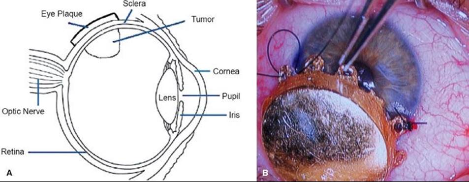
Plaque Brachytherapy
Plaque brachytherapy is the mainstay of treatment in many centers, with iodine-125 and ruthenium-106 being the most common isotopes, although palladium-103 has also been used effectively.91 Iodine emits γ-rays, which have a range sufficient for tumors up to 8- to 10-mm thick, while ruthenium delivers beta-particles that have a more limited range, which is suitable for tumors up to approximately 5 mm. The general objective with all plaques is to deliver approximately 80 Gy to the tumor apex by fixing the plaque in the exact location of the tumor (Fig. 38.6). Computer modeling has been developed, as seen in general radiotherapy planning, to create a three-dimensional model of the eye and determine the appropriate treatment time and estimated dose to the optic nerve, macula, and lens.92 The largest prospective experience of patients treated by plaque brachytherapy was conducted by the Collaborative Ocular Melanoma Study (COMS) group. From 1986 to 2003, COMS conducted two multicenter trials of brachytherapy with iodine-125 versus enucleation in selected patients with choroidal melanoma. Long-term results were subsequently published in COMS report 28 in 2006, which evaluated 1,317 patients.93 Eligible patients had unilateral choroidal melanoma with an apical height of 2.5 to 10 mm and a maximum basal tumor diameter (MBTD) of 16 mm. Patients whose tumors were contiguous with the optic disc were ineligible, as were patients with metastases from melanoma or another malignancy. Patients were followed for 5 to 15 years, and within 12 years after enrollment, 471 of 1,317 (36%) had died. Overall cause-specific mortality at 5 and 10 years for both treatment arms were 19% and 35%, respectively. Cumulative all-cause mortality was 43% in the iodine-125 arm and 41% in the enucleation arm, indicating that with long-term follow-up no survival difference existed between plaque brachytherapy and enucleation. Age older than 60 years and MBTD >11 mm were the primary predictors of time to death from all causes and death with melanoma metastases. Local control was excellent in the COMS series, with only 12.5% of patients requiring salvage enucleation.94 Visual acuity after plaque brachytherapy depends on a number of features. Shields et al.95examined 1,106 patients with visual acuity of 20/100 or better who underwent brachytherapy for uveal melanoma. They found 34% of patients had poor visual acuity at 5 years and 68% at 10 years, defined as 20/200 to no light perception. Factors adversely affecting visual acuity were age over 60 years, poor vision at baseline, increasing tumor thickness (>8 mm), proximity to the foveola of <5 mm, recurrent tumor, subretinal fluid, and history of diabetes or hypertension. Best results were obtained in eyes with small tumors outside a radius of 5 mm from the optic disc and foveola. Whether improvements in tumor imaging and localization and radiation planning will improve functional results will be determined with further investigation.
FIGURE 38.6. A: Eye anatomy and plaque placement in location of tumor. (From Chaudhari S, Deshpande S, Anand V, et al. Dosimetry and treatment planning of Occu-Prosta I-125 seeds for intraocular lesions. J Med Phys 2008;33:14–18.) B: Plaque brachytherapy for melanoma of the iris. (From Khan MK, Khan N, Almasan A, et al. Future of radiation therapy for malignant melanoma in an era of newer, more effective biological agents. Onco Targets Ther 2011;4:137–148.)
Proton Beam Therapy
Proton beam therapy may be used to treat uveal melanoma and is usually indicated for tumors that extend close to or are contiguous with the optic disc, referred to as parapapillary and peripapillary tumors, respectively.96 Large tumors and lesions of the iris and ciliary body may also be candidates for this modality as are patients who are not fit for operative therapy.79 Because a proton beam delivers a homogeneous dose to tumor and has a sharp edge, a high tumor dose can be delivered with relative sparing of the optic nerve. The decision to use proton therapy over other forms of external therapy, such as helium ions or stereotactic radiotherapy, often depends on the availability of treatment facilities in addition to clinical factors. One of the largest experiences in proton beam therapy for uveal melanoma is from the Harvard Medical School.97 Treatment planning involves intraoperative examination by transillumination or indirect ophthalmoscopy, and the edges of the tumor are delineated by four tantalum rings sutured to the sclera. For tumors of the ciliary body and peripheral choroid, surgery is not performed; transillumination is instead used to define tumor margins in relation to the iris and cornea. Radiation planning is done with an interactive three-dimensional computer system to define the beam aperture and range modulation needed to adequately encompass the tumor and a 1.5-mm margin is included to allow for motion during treatment, setup error, and possible microscopic extension.98 Patients receive a total dose of 70 CGE, which is delivered in 5 equal fractions over 7 to 10 days (63.6 proton Gy é 1.1 relative biological effectiveness = 70 CGE).97 The Harvard group recently reported long-term follow-up in a series of 573 patients with peripapillary and parapapillary melanomas with median surveillance of 96.3 months.96 Local recurrence was rare, with 5- and 10-year local recurrence of 3.3% and 6%, respectively, and similarly high rates of local control have been observed in a larger series from the same institution.99Enucleation rates were 13.3% and 17.1% at 5 and 10 years after treatment, respectively. Of 450 patients with baseline visual acuity of 20/200 or better, two-thirds had visual acuity <20/200 years after treatment, although 56% could count fingers. The most common complications were radiation maculopathy and papillopathy. By 3 years the cumulative rate of both complications was 49%, and by 10 years, this increased to 61% for papillopathy and 68% for maculopathy. The visual outcome after proton therapy depends on the height of the tumor and its location relative to the fovea and optic nerve.99 Anterior segment complications, such as rubeosis iridis and neovascular glaucoma, are the most serious, but occur less frequently, with each occurring in approximately 15% of cases at 5 years postradiation.100 These results indicate that although visual acuity is compromised with proton beam therapy, particularly in patients with juxtapapillary lesions, some preservation is possible and eye conservation is likely with extremely low rates of local recurrence.
Stereotactic Radiotherapy
Stereotactic radiation can be delivered by linear accelerator (LINAC) or by specialized devices for focused radiation such as the Leksell Gamma Knife (Elekta, Norcross, GA), which provides focused radiation with a multitude of sources. Because gamma knife treatment is usually done in a single fraction, the term radiosurgery is applied, whereas for LINAC-based treatment, single fraction or multifraction treatment is possible. Gamma knife radiosurgery (GKR) for uveal melanoma was first introduced in 1998 by investigators at the Indiana University School of Medicine.101 Nineteen patients with uveal melanoma were treated to a dose of 40 Gy prescribed to the 50% isodose line. With median follow-up was 40 months, 3- and 5-year overall survival rates were 86 and 55%, respectively. The 3- and 5-year tumor control rates were both 94%. Six of the 19 treated patients (32%) developed distant metastasis 31 to 75 months after GKR. Of the 19 patients treated, 2 had improved, 4 had stable, and 13 had worse vision in the treated eye. Similar results were reported in a larger series of 78 patients from investigators in Milan.102 The dose was adjusted over the treatment period: 7 patients received 50 Gy at the 50% isodose line (1994–1995), 21 patients received 40 Gy to the 50% isodose line (1995–1999), and 47 patients received 35 Gy to the 50% isodose line (2000–2006). Local tumor control was achieved in 91.0% of patients and the eye retention rate was 89.7%. A significant relative reduction of visual acuity was observed during follow-up. The most frequently encountered complications were exudative retinopathy (33.3%), neovascular glaucoma (18.7%), radiogenic retinopathy (13.5%) and vitreous hemorrhage (10.4%).
One theoretical disadvantage of single-dose stereotactic radiosurgery is the potential for complications, which has prompted investigators to explore fractionated stereotactic radiotherapy (SRT) treatment of uveal melanoma.103 In particular, severe radiation retinopathy is dose dependent with respect to both total dose (>25 Gy) and dose per fraction (>2 Gy).104,105 Muller et al.103 conducted a prospective study on 102 patients with uveal melanoma treated with fractionated SRT between 1999 and 2007. Patients had uveal melanoma of the choroid or ciliary body with a tumor thickness <12 mm and diameter <16 mm with no metastases. The technique, like GKR, utilized a fixed immobilization system, and a dose of 50 Gy was delivered in 5 equal fractions on 5 consecutive days using 6 MV photons with stereotactic arcs. With median follow-up of 32 months, local control was achieved in 96% of patients. Fifteen enucleations were performed 2 to 85 months after radiation, and best corrected visual acuity (defined as 20/é while using glasses if needed) decreased from a mean of 0.26 at diagnosis to 0.16, 3 months after radiation and then declined to 0.03, 4 years after therapy. Deterioration of visual acuity was in part related to complications of treatment. Grade 3 or 4 neurovascular glaucoma occurred in 9 patients, 8 of which required enucleation. In these 9 patients, tumors were not anteriorly located nor did they receive a high dose to the ciliary body, as one might expect, but were associated with grade 3 retinopathy, which was seen in 19 patients. The authors hypothesized that the physical reaction of ischemic retinopathy might also affect vessels in the ciliary body. Thirteen patients (13%) developed grade 3 or 4 optic neuropathy, which was associated with posterior tumors and optic nerve dose, which was limited to 4 Gy per fraction. Grade 3 cataracts occurred in 10 patients (10%), which were managed by extraction and lens implantation. Cataract formation was dose related, with a median dose of 5 Gy per fraction to the lens, causing cataract in 50% of cases. The authors concluded that while local control was excellent, the number of secondary enucleations was substantial, particularly from neurovascular glaucoma. These data suggest that further follow-up is needed to determine response durability and late side effects of this treatment program.
Retinoblastoma
Retinoblastoma (Rb), the most common ocular malignancy in childhood, affects approximately 300 children per year in the United States.104 The incidence is higher in developing countries, and while the reason for this is not clear, lower socioeconomic status and the presence of certain human papilloma virus sequences have been implicated.105 Rb has a heritable form and a nonheritable form, with approximately 55% of children having the nonheritable form. If there is no family history, the disease is labeled sporadic, but this does not necessarily indicate that it is the nonheritable form, because bilateral cases, most of which are heritable, often have no family history of Rb. It can present with unilateral disease (two-thirds of cases), bilateral disease, or rarely with tumor in both eyes and the pineal gland, which is called trilateral disease.106 Approximately 80% of children in Rb are diagnosed before the age of 3, with unilateral cases diagnosed at an earlier age (14–16 months) than those with bilateral presentations (29–30 months).107–110 Histologically, Rb develops from immature retinal cells and replaces the retina and other intraocular tissues. Macroscopically, viable tumor cells are found near blood vessels, while zones of necrosis are found in relatively avascular areas. Microscopically, both undifferentiated and differentiated elements may be present. Undifferentiated elements appear as collections of small, round cells with hyperchromatic nuclei; differentiated elements include Flexner-Wintersteiner rosettes, Homer-Wright rosettes, and fluerettes from photoreceptor differentiation.111
The study of Rb has provided insights into the genetic basis of cancer. The Rb gene (RB1) is located on the long arm of chromosome 13 (13q14). In order for Rb to develop, both copies of the gene at the 13q14 locus must be lost, deleted, mutated, or inactivated. If either the maternal or paternal copy of the gene that is inherited by an individual is defective, then that individual is heterozygous for the mutant allele. Tumor formation requires both alleles of the gene to be mutant or inactive. These two mutations correlate to the two “hits” theorized by Knudson112 and Hethcote and Knudson,113 which was based on the finding that children with bilateral Rb developed multifocal, bilateral tumors at an earlier age than those with unifocal, unilateral tumors. The first “hit” can be inherited and would be present in all cells in the body, and the second “hit” results in loss of the remaining normal allele and occurs within a particular retinal cell or cells with dysregulation of the cell cycle.114 In sporadic, nonheritable Rb, both hits occur within a single retinal cell after fertilization (somatic events), thus resulting in unilateral Rb. Identifying the RB1mutation can have management implications both in the affected child as well as siblings and future offspring. For example, if RB1 mutation is detected, then siblings, children, and other relatives can be tested for the mutation. If they do not carry the mutation, they need not undergo rigorous examinations under anesthesia.115
The most common and obvious sign of Rb is leukokoria, a discoloration of the pupil (Fig. 38.7). Other less common and less specific signs and symptoms are deterioration of vision, a red or irritated eye, faltering growth, or developmental delay.116 Some children with retinoblastoma can develop a squint, commonly referred to as cross-eyed or wall-eyed, indicating strabismus.117 In advanced disease in developing countries, eye enlargement is a common finding. Funduscopy typically reveals a white-colored main tumor (Fig. 38.8), frequently with satellite lesions in the retina, subretinal space, or vitreous referred to as “seeds.” Secondary serous retinal detachment may be associated with large lesions. To confirm these findings, a detailed examination under anesthesia through dilated pupils is performed.
Ultrasonography of the eyes is often performed to evaluate the intraocular mass with attention to heterogeneity and calcifications, which support a diagnosis of Rb. Ultrasonography is not as sensitive as CT, which is the ideal imaging format to detect intraocular calcifications. CT, however, raises the concern of exposure to radiation in children younger than 1 year of age with germline mutations,118 but it is still frequently used to confirm the diagnosis. Magnetic resonance imaging (MRI) of the brain and orbits is the most sensitive means of evaluating for extraocular extension and also provides better delineation of the optic nerve and the pineal area.119 MRI of the brain and spinal cord and cerebral spinal fluid examination are indicated when there is gross invasion of the optic nerve by imaging studies or microscopic involvement beyond the lamina cribrosa on histopathologic examination of the enucleated eye. A bone marrow examination and a bone scan are indicated only in cases of an abnormal blood count or clinical symptoms suggesting osseous metastases. The diagnosis of retinoblastoma is based on examination by an ophthalmologist and imaging studies. Biopsy is generally not performed due to the theoretical risk for extraocular dissemination, which could convert an intraocular, curable tumor into extraocular, metastatic disease. Therefore, in the absence of a tissue diagnosis, benign conditions that can resemble retinoblastoma must be carefully excluded.
Rb can spread in a variety of ways, including direct invasion of the optic nerve into the chiasm or dissemination through the subarachnoid space to the brain and spinal cord. Tumors can also invade the choroid and the vascular layer and spread via blood to the bone and bone marrow.120,121 Anterior spread can occur and involve the aqueous venous channels, conjunctiva, and lymphatics or invade the sclera into the orbit with eventual spread to regional lymph nodes.
FIGURE 38.7. Leukokoria from retinoblastoma. (From Aerts I, Lumbroso-Le Rouic L, Gauthier-Villars M, et al. Retinoblastoma. Orphanet J Rare Dis 2006;1:31.)

FIGURE 38.8. Retinoblastoma on fundoscopic exam. (From Aerts I, Lumbroso-Le Rouic L, Gauthier-Villars M, et al. Retinoblastoma. Orphanet J Rare Dis 2006;1:31.)

Treatment
Management of Rb requires close cooperation of a multidisciplinary team of ophthalmologists, pediatric oncologists, pediatric radiation oncologists, pathologists, genetic counselors, social workers, nurses, and others. Most unilateral cases present with advanced intraocular disease and require enucleation, which is also indicated for eyes with recurrent disease and no useful vision. Children with bilateral presentations typically require multimodality therapy with chemotherapy and local therapy. Staging and classification schemes have been developed to assess outcomes, particularly for intraocular Rb. The Reese-Ellsworth (R-E) classification, developed in the 1960s, was the first of these (Table 38.2).122 Specifically, the R-E classification, which has five groups, was devised to predict prognosis in eyes that were treated with EBRT. Eyes with disease consistent with the lower groups have a lower risk for enucleation following EBRT, while group V eyes have the highest risk for enucleation. In recent years, chemotherapy has diminished the role EBRT for intraocular disease, and thus the R-E scheme is less useful and alternative schemes have been proposed by Shields et at.123 and Murphree,124 among others. The International Retinoblastoma Classification was developed by a group of experts in 2003 in Paris, which is used by many cooperative study groups (Table 38.3). An additional system has been proposed by Chantada et al.,125 which was designed to address extraocular disease and microscopic disease following enucleation.
TABLE 38.2 REESE-ELLSWORTH CLASSIFICATION OF RETINOBLASTOMA

TABLE 38.3 INTERNATIONAL CLASSIFICATION OF RETINOBLASTOMA

Enucleation
Most children with unilateral Rb present with advanced disease and require enucleation. Other indications for enucleation are cases of bilateral disease where the eye with more advanced disease does not respond to chemotherapy or other treatments, when active tumor is present in an eye with no vision, when glaucoma is present as a result of neovascularization of the iris or tumor invasion into the anterior chamber, and when direct visualization of an active tumor is obstructed by conditions including hemorrhage, corneal opacity, or cataract.126 Enucleation is curative in >95% of patients with unilateral disease. Orbital implants are used at most treatment centers, and by connecting them to orbital muscles, excellent cosmesis can be achieved.
External Beam Radiation Therapy
Rb is a highly radiosensitive tumor as first shown by Hilgartner127 in 1903. By virtue of its radiosensitive nature, Rb was historically treated with first-line EBRT in a majority of cases, with doses of 42 to 50 Gy given in 1.5- to 2-Gy fractions, resulting in 87% eye preservation in R-E groups I to IV versus 29% in group V eyes.128 However during the past 15 years, the trend has been to move away from radiotherapy because of radiation-induced growth deformities of the bony orbit and mortality related to osteosarcoma development.129–131 However, EBRT still has a viable role in the management of Rb, particularly as salvage, or perhaps more accurately termed, consolidative treatment in tumors that are refractory to chemotherapy and other local therapies. Other indications include lesions that are too large, numerous, or close to the optic disc or fovea for focal therapy out of concern for preserving central vision. EBRT also has a special role in treating eyes with vitreous seeds.132 Chan et al.133 reported on 36 eyes that received EBRT after incomplete response to primary chemotherapy and focal therapies in 22 patients with bilateral Rb. Thirty-two received lens-sparing EBRT and the remainder received whole-eye EBRT to a dose of 40 to 44 Gy in 20 to 22 fractions. The rate of eye preservation was 83%, and 67% required no further treatment after EBRT. Visual acuity was recorded for 19 eyes, of which 10 read 6/9-6/5, 3 read 6-18-6/36, and 6 read 6/60 or worse. Side effects were limited to cataracts and dry eyes, although follow-up (median 40 months) was insufficient to assess second malignancies. The investigators concluded that EBRT was highly effective in preserving eyes with useful vision in bilateral Rb in cases refractory to chemotherapy and focal therapies. In a more advanced population, Kingston et al.134 evaluated consolidative EBRT (40–44 Gy) in 14 patients with R-E group V eyes after induction chemotherapy with carboplatin, etoposide, and vincristine. Four eyes were enucleated primarily for severe disease at presentation and of the remaining 20, 6 required enucleation (4 for recurrence, 2 for neovascular glaucoma). Of the 12 surviving children, 5 have visual acuity of better than 1/60 in at least one eye. Thus, while most group V eyes could be salvaged, the resultant visual acuity was often poor.
Radiation techniques in Rb should provide uniform coverage of the entire retina approaching the ora serrata and coverage of up to 10 mm of the optic nerve while sparing the lens and bony anatomy to the extent possible.132 In practice, the dual goals of including the entire retina in the beam and protecting the lens have represented a challenge. Historically, a single lateral field was used in the radiotherapeutic management of one eye and parallel-opposed fields for the management of both eyes. The traditional border for these “D-shaped” fields was the lateral rim of the bony orbit, which could result in underdosing of the anterior retina with associated local failure. McCormick et al.135 reported local failure in two-thirds of cases when lens-sparing lateral technique was used. Based on these concerns, more sophisticated techniques were developed, such as the anterior lens-sparing technique, modified lateral field techniques using oblique angles, anterior treatment techniques using electrons, and multiple noncoplanar arcs.136–139
Conformal or IMRT is well suited for the treatment of small tumors such as Rb. Krasin et al.140 performed a planning study that favored the use of IMRT over conformal, anterior-lateral photon and anterior electron plans for the treatment of the entire globe. IMRT resulted in the greatest sparing of the surrounding bony orbit and other normal tissues. As acute and late toxicity is related to volume and dose of normal tissue irradiated, proton therapy has been considered for the treatment of Rb, with the added advantage of reducing low-dose irradiation of normal tissue. Lee et al.141 compared protons with three-dimensional conformal radiation therapy, IMRT, and electron therapy when treating the entire retina and vitreous cavity. Protons provided superior coverage with greater sparing of normal tissue, which in theory may result in a superior therapeutic ratio. Krengli et al.142 came to a similar conclusion looking at target coverage and lens sparing using protons for various intraocular tumor locations and beam arrangements. Focused techniques such as proton beam irradiation should be strongly considered in the management of Rb due to the high risk of second malignancy. Patients with hereditary disease who received EBRT have a cumulative incidence of second cancers of 35%, compared with 6% for those who did not receive EBRT, and the risk is even higher if treatment takes place before 1 year of age.143,144 Cataracts, optic nerve damage, total retinal vascular occlusion, vitreous hemorrhage, and facial and temporal bone hypoplasia are other complications associated with EBRT therapy.145
Brachytherapy
Brachytherapy with either iodine-125, gold, and more recently ruthenium have been used in selected cases of Rb.146 The intention is to deliver a dose of 40 to 45 Gy transclerally to the apex of the tumor over a period of 2 to 4 days. This treatment is limited to tumors that are <16 mm in base and 8 mm in thickness, and can be used as the primary treatment or, more frequently, in patients who had failed initial therapy, including previous EBRT.147–150 Shields et al.151 described 79% local control at 5 years using this method. Side effects are generally less common than with EBRT and include optic neuropathy, radiation retinopathy, and cataract formation. Second malignancies do not appear to be associated with this type of local therapy. Relative contraindications include larger tumors and those that involve the macula.
Thermotherapy
Thermotherapy involves the application of heat directly to the tumor with infrared radiation. A temperature between 45°C and 60°C is reached, which is below the coagulative threshold and therefore spares the retinal vessels from coagulation.151,152 Thermotherapy alone can be used for small lesions that are ≤3 mm in diameter without vitreous or subretinal seeds. In a study of 91 tumors, 92% of the tumors that were <1.5 mm in diameter were controlled with thermotherapy alone.153
Chemothermotherapy and Laser Photocoagulation
Larger tumors or tumors with subretinal seeds are usually treated with a combination of thermotherapy and chemotherapy (chemothermotherapy), usually delivered within hours of each other. In one study of 188 retinoblastomas, tumor control was achieved in 86% of cases.154 Complications included focal iris atrophy, paraxial lens opacity, sector optic disk atrophy, retinal traction, optic disk edema, retinal vascular occlusion, retinal detachment, and corneal edema. Chemothermotherapy may be especially useful for patients with small tumors adjacent to the fovea and optic nerve, where radiation therapy or laser photocoagulation may result in significant visual loss. Laser photocoagulation is recommended only for small posterior tumors with the goal of coagulating the tumor’s blood supply.155 Effective therapy usually requires two or three sessions at monthly intervals. Complications of this treatment include retinal detachment, retinal vascular occlusion, retinal traction, and preretinal fibrosis.
Cryotherapy
Cryotherapy induces tumor tissue to freeze rapidly, resulting in damage to the vascular endothelium with secondary thrombosis and infarction of the tumor tissue. It may be used as primary therapy for small peripheral tumors or for small recurrent tumors previously treated with other modalities. Tumors are typically treated three times per session, with one or two sessions at monthly intervals. Ninety percent of tumors <3 mm in diameter are cured permanently, and complications are few and rarely serious.156 Transient conjunctival edema and transient localized serous retinal detachments can occur. Vitreous hemorrhage can be observed in large or previously irradiated tumors.
Chemotherapy
Chemotherapy has been used to treat intraocular retinoblastoma since the early 1990s. Chemotherapy is used to reduce the size of the tumor to allow local ophthalmological therapies, including cryotherapy and laser photocoagulation, or thermotherapy, to eradicate the remaining disease. This combination of therapies has been promoted to avoid EBRT or enucleation and thereby decreases the potential for long-term side effects while salvaging useful vision. The common indications for chemotherapy for intraocular Rb include tumors that cannot be effectively treated with local therapies alone, usually due to size. Chemotherapy may be indicated in children with unilateral or bilateral disease, but many patients with unilateral disease are diagnosed with advanced disease and require enucleation. Numerous studies have been published that show that chemotherapy is very effective in eliminating the need for EBRT or enucleation in R-E group I to III eyes, while proving to be significantly less successful in eyes with group IV or V disease.157–161,162,163–165 Carboplatin, vincristine, and etoposide are generally used, and cyclosporine has been added to the regimen in some institutions in order to reduce drug resistance.166 Chemotherapy regimens from different investigators vary in the number and frequency of cycles, but they are generally well tolerated, with the expected side effects of myelosuppression and associated risk of infection. There is still the potential risk for second malignancies, especially when using etoposide.167 Chemotherapy alone is not very effective in avoiding EBRT or enucleation in patients with R-E group V eyes, especially those with vitreous seeds. Friedman et al.165 showed that only 53% of 30 group V eyes could be controlled with chemotherapy alone. Based on these data, newer regimens incorporate carboplatin, vincristine, and etoposide along with subtenon carboplatin for more advanced eyes.168,169 Eyes with diffuse vitreous seeding rarely respond to chemotherapy alone, and while EBRT is modestly successful, new approaches are needed. Novel therapies for patient with vitreous or subretinal seeding include intra-arterial chemotherapy (IAC) with selective catheterization of the ophthalmic artery.170 Gobin et al.170 described 78 patients (95 eyes) treated with IAC with melphalan with or without topotecan and evaluated procedure success, event-free (RT or enucleation) ocular survival, and ocular and extraocular complications. The procedure succeeded in 98.5% of cases and the ocular event-free survival rates at 2 years were 70% for all eyes, 82% for eyes that received IAC as primary treatment, and 58% for eyes that had prior treatment with chemotherapy or EBRT, and there were no permanent complications. Abramson et al.171 reported similarly encouraging results in 67 patients (76 eyes) treated with IAC. Among treatment-naïve eyes, the ocular salvage rate was 83% for eyes with subretinal seeding only, 64% for eyes with vitreous seeding only, and 80% for eyes with both. Other strategies have also been investigated. A recent phase I study using adenoviral vectors to deliver the herpes simplex thymidine kinase gene followed by ganciclovir demonstrated durable clinical and histopathologic responses in patients heavily pretreated with vitreous seeds.172 Clinical trials using these novel therapies should be encouraged in this population.
Management of Extraocular Disease
Patients with extraocular disease historically have had a poor prognosis, but recent studies using combinations of chemotherapy and EBRT have been encouraging. Chantada et al.173 reported a 5-year event-free survival rate of 84% in 15 patients with orbital or preauricular disease treated with chemotherapy that included vincristine, doxorubicin, and cyclophosphamide or vincristine, idarubicin, cyclophosphamide, carboplatin, and etoposide. These patients also received EBRT of 45 Gy administered to the optic chiasm for patients with orbital disease and to the involved nodes for those with preauricular lymphadenopathy. There are several reports suggesting that high-dose chemotherapy with stem cell rescue combined with EBRT for areas of bulky disease at diagnosis is beneficial, with some long-term survivors among patients with metastatic disease not involving the central nervous system.174–176,177
Primary Intraocular Lymphoma
Primary intraocular lymphoma (PIOL), formally known as ocular reticulum cell sarcoma, is an uncommon clinical manifestation of non-Hodgkin lymphoma, which arises in the retina or the vitreous humor.178 It usually develops in patients in the fifth and sixth decades of life as a chronic, relapsing, and steroid-resistant uveitis and vitritis. Patients often complain of blurred vision, a painless loss of vision, and floaters. Intraocular lymphoma may occur independently, prior or subsequent to a primary central nervous system lymphoma. PIOL progresses to intracranial involvement in 60% to 85% of cases.179,180 Historically PIOL has been difficult to diagnose, often taking several years from the onset of symptoms to establishing a diagnosis, which likely contributed to suboptimal outcome in many cases.181 Given the tumor’s rarity, the optimal treatment of PIOL is unclear. Radiation therapy has been used with durable control after 35 to 45 Gy given exclusively to both eyes in the absence of central nervous system disease.179,182 However, concerns of central nervous system relapse have led investigators to use chemotherapy as initial treatment and radiation as a consolidative therapy.183,184 Methotrexate and cytosine arabinoside are most commonly used, given their ability to cross the blood–ocular barrier.
Optic Pathway Glioma
Optic pathway gliomas, with or without contiguous involvement of the hypothalamus, have an incidence of approximately 1 in 100,000, with 90% presenting in the first two decades of life185 Most of these neoplasms are pilocytic or low-grade astrocytomas.186 Untreated, the clinical course is that of deterioration of visual acuity, progressive visual field deficits, endocrine or intellectual impairment, and death in up to 30% due to local tumor progression.187,188Multiple treatment strategies exist, including surveillance, chemotherapy, radiation therapy, surgery, or some combination of therapies. Advances in management have resulted in cause-specific survival rates of 90% to 100% except in cases associated with neurofibromatosis, which carry a worse prognosis.189,190 Therefore, multidisciplinary management of optic pathway gliomas is critical with an emphasis on reducing treatment-related sequelae. The role of surgery is limited but may be a reasonable treatment option in patients where tumor is confined to a single optic nerve with no useful vision. Local failure rates of approximately 5% can be achieved after complete resection.185Radiation therapy has resulted in 10-year survival rates ranging from 40% to 93%, but with potentially severe long-term sequelae, including endocrine problems, neurodevelopmental disorders, and second malignancy.191 Concerns of these toxicities are significant enough such that at most centers, children of any age are initially treated with chemotherapy in order to delay irradiation until progression. Investigators have found that a 2.5- to 3-year delay in RT can be achieved with this approach.189
The main indication of RT is for progressive disease, and radiation doses in the range from 45 to 60 Gy in 1.8- to 2.0-Gy fractions have been effective.190,192 Erkal et al.190 reported on 33 cases of optic pathway gliomas (OPG) treated between 1973 and 1994. Twenty-four children had OPGs and nine had chiasmatic-hypothalamic gliomas. Evidence of neurofibromatosis was present in six children. Subtotal resection was performed in 22 and biopsy in 7. Median total dose was 50 Gy in 25 fractions and mean follow-up was 13.6 years. Ten-year overall, progression-free, and cause-specific survival rates were 79%, 77%, and 88%, respectively. Differences in any of the survival endpoints between optic pathway and chiasmatic-hypothalamic gliomas were not statistically significant, but absence of neurofibromatosis correlated with significantly better progression-free and cause-specific survival. Grabenbauer et al.192 also reported a series of OPG but also assessed visual outcomes. Twenty-five patients received radiation therapy following surgery or biopsy. Treatment volume included a 0.5- to 1-cm margin around the tumor as depicted on CT or MRI. Age-adjusted radiation doses ranged from 45 to 60 Gy with a fraction size of 1.6 to 2 Gy. Overall survival and progression-free survival rates were 94% and 69% at 10 years, respectively. Age older than 10 years at time of treatment and total radiation dose >45 Gy significantly improved progression-free survival. Hypothalamic deficiency also correlated with age, with 69% patients aged below 10 years compared with 25% above 10 years experiencing this condition. As for visual acuity, 36% had an improvement, 52% remained stable, and 12% had measurable deterioration. The authors concluded that postoperative RT with a total dose above 45 Gy should be considered for patients with progressive OPG. Lifelong yearly evaluation for growth hormone, thyroid, and adrenal function is also crucial because the need for replacement therapy is high, particularly in children under 10 years of age.
As in other pediatric malignancies, radiation technique should be approached with an emphasis on normal tissue sparing. Combs et al.193 reported on 15 patients treated with fractionated stereotactic RT (FSRT) to a median total dose of 50.2 Gy at 1.8 Gy per fraction. The progression-free survival rate at 3 and 5 years was 92% and 72%, respectively. Functional results were encouraging, with only two patients having worsening of vision after RT. Seven had preexisting endocrinopathy, but only one additional patient had endocrine dysfunction after treatment, suggesting FSRT is an effective option for OPG. Still, conformal RT, IMRT, and FSRT carry concerns of second malignancy due the large volume of intracranial tissue receiving a low, potentially mutagenic dose of radiation. For this reason, proton beam therapy has been advocated.194 Fuss et al.194 evaluated dosimetry plans for proton versus conformal plans in seven cases of OPG and found proton therapy offered substantial normal tissue sparing both at high- and low-dose areas. The difference was more apparent in tumors >80 cm3, but even in tumors <20 cm3, conformity of conformal RT came at the expense of a larger amount of normal tissue receiving low to moderate doses of radiation.
Orbital Tumors
Primary Orbital Lymphoma
Orbital non-Hodgkin lymphomas (NHL) account for only 0.01% of NHL and are typically B-cell lymphomas.195 Depending on the stage and grade of disease, observation, first-line chemotherapy, or chemotherapy followed by RT are all treatment options, but RT is generally the treatment of choice for NHL localized to the orbital cavity.196,197 Local control at the rate of 90% to 100% can be achieved using doses of 30 to 36 Gy.197 Historically, radiation was delivered with an anterior portal prescribed to the orbital apex or by wedged anterior and later fields. Late effects from these techniques include cataract formation, lens ulceration, glaucoma, and lacrimal complications.197 More recently, investigators have described the use of IMRT in orbital lymphoma and were able to reduce dose to the contralateral orbit, lacrimal gland, and lens (Fig. 38.9).198
Conjunctival Tumors
Conjunctival tumors comprise a variety of conditions, from benign papilloma to malignant lesions such as squamous cell carcinoma (SCC). Radiotherapeutic management of these tumors is related to the histologic type, as is the case in tumors found elsewhere. Mucosa-associated lymphoid tissue (MALT) lymphoma, for example, has been effectively managed with exclusive RT with local control rates approaching 100%.199 In a series of patients with MALT lymphoma of the ocular adnexa that included 37 patients with conjunctival disease, Hashimoto et al.200 reported local control rates of 100% with a median dose of 30.6 Gy. Electron beam therapy was frequently used with eye lens shielding when possible depending on the extent of disease (Fig. 38.10). Treatment options for SCC include excision with or without adjuvant cryotherapy or topical chemotherapy, while advanced cases may require orbital exenteration.201,202 RT has been used in the primary, adjuvant, and salvage settings with a variety of methods, including photon, proton, and electron beam therapy.203,204 Most frequently, however, radiation is used in patients with relapsed disease. The treatment volume typically includes gross tumor with margins of approximately 1 cm and doses of approximately 60 Gy, as in cases of SCC from other sites.205
FIGURE 38.9. Primary orbital lymphoma treated with intensity modulated radiation therapy illustrating sparing of lens, lacrimal gland, and contralateral orbit.

FIGURE 38.10. Electron beam therapy for mucosa-associated lymphoid tissue lymphoma of the conjunctiva. The patient received 30.6 Gy using 9 MeV electrons. Lens shielding was not used due to the extent of disease at presentation.

FIGURE 38.11. A: Orbital rhabdomyosarcoma with proptosis. B: Coronal computed tomography scan of patient with orbital rhabdomyosarcoma. (From Das JK, Tiwary BK, Paul SB, et al. Primary orbital rhabdomyosarcoma with skeletal muscle metastasis. Oman J Ophthalmol2010;3:91–93.)

Sebaceous Carcinoma of the Eyelid
Sebaceous gland adenocarcinoma occurs in the periorbital area, usually in the eyelid.206 It can exhibit aggressive local behavior and can metastasize to regional lymph nodes and distant organs. Older individuals tend to be affected, but it occurs with greater frequency and at an earlier age in patients with hereditary retinoblastoma with treated with radiation.207–209 The main systemic association is Muir-Torre syndrome, an autosomal dominant condition characterized by greater frequency of sebaceous adenoma, sebaceous carcinoma, keratoacanthoma, and gastrointestinal malignancies.210 The most common method of metastasis of eyelid sebaceous carcinoma is through the lymphatic channels to regional lymph nodes. Historically, regional node metastasis occurred in about 30% of cases, but metastases have become less frequent in recent years.211,212 Tumors that originate in the upper eyelid tend to metastasize to preauricular and parotid nodes, which represent the most common sites of metastasis. Tumors of the lower eyelid region can metastasize to submandibular and cervical nodes, which warrant consideration in RT planning. Until recently, orbital exenteration was widely believed to be the only reasonable option in the management of sebaceous carcinoma that involved most of the conjunctiva and invaded the orbit. However, local excision with adjuvant therapies, such as topical chemotherapy or cryotherapy, have become more accepted.213,214 EBRT, with doses of approximately 60 Gy, is rarely used for primary treatment, but should be considered for adverse features such as regional lymph node involvement or for recurrent disease.215
Orbital Rhabdomyosarcoma
Rhabdomyosarcoma (RMS) affects 250 to 300 children per year in the United States. In 1950, Stobbe and Dargeon216 demonstrated improvement in the outcome in head and neck sites when radiation therapy was added after incompletely resected RMS. In 1961, Pinkel and Pinkren217 advocated adjuvant chemotherapy after complete surgical excision and postoperative radiation therapy, which was the beginning of the multimodal approach to solid tumors. Recognizing the value of this multimodal approach as well as the relative rarity of these tumors, the leadership of the three U.S. cooperative pediatric cancer research groups formed the Intergroup Rhabdomyosarcoma Study (IRS) group in 1972 to investigate the biology and treatment of RMS. Since then, five successive clinical protocols involving almost 5,000 patients have been completed: IRS-I (1972–1978); IRS-II (1978–1984); IRS-III (1984–1991); IRS-IV Pilot (for patients with advanced disease only; 1987–1991); and IRS-IV (1991–1997).218,219–223 The head and neck region is the most common site of presentation and accounts for about 35% of the patients in the IRS studies.224 These tumors are most commonly of the embryonal subtype and rarely spread to regional lymph nodes. Orbital tumors produce proptosis and, occasionally, ophthalmoplegia (Fig. 38.11). Modern management of RMS comprises multiagent chemotherapy followed by RT. Prechemotherapy as well as postchemotherapy tumor volume must be considered when planning RT. McDonald et al.225 reported on 20 patients with head and neck RMS (two orbit) treated with IMRT to a total dose of 50 Gy, in accordance with cooperative group protocols. The initial targeting of the prechemotherapy tumor volume with 1- to 2-cm margin to 30.6 to 36 Gy followed by a cone-down boost to the postchemotherapy tumor volume with a 0.5- to 1-cm margin allowed for significant sparing of normal tissues and only one patient developed an in-field recurrence after 50 months. The 3-year event-free survival, overall survival, and risk of central nervous system failure were 74%, 76%, and 7%, respectively.
Periorbital Skin Cancers
Skin cancers of the eyelid and periorbital skin include, most frequently, basal cell carcinoma (BCC), SCC, but also rare histologies such as Merkel cell carcinoma (MCC).203,226 For small BCC and SCC, single-modality treatment with surgery or RT results in good local control, but RT is frequently used due to cosmetic concerns. For more advanced tumors or more aggressive histologies, such as MCC, combined surgery and RT is typically recommended. Low energy electron beam therapy is useful in this setting and can be delivered with an eye shield. RT for skin tumors requires doses of 60 Gy or more for definitive treatment using standard fractionation, but altered fraction can be employed to shorten treatment duration and minimize ocular trauma of shield placement.
![]() REFERENCES
REFERENCES
1. American Cancer Society. Eye cancer (melanoma and lymphoma). Available at: http://www.cancer.org/Cancer/EyeCancer/DetailedGuide/eye-cancer-key-statistics.
2. Shields CL, Shields JA, Gross N, et al. Survey of 520 eyes with uveal metastases. Ophthalmology 1997;104:1265–1276.
3. Karp LA, Streeten BW, Cogan DG. Radiation-induced atrophy of the Meibomian gland. Arch Ophthalmol 1979;97:303–305.
4. Stephens LC, Schultheiss TE, Peters LJ, et al. Acute radiation injury of ocular adnexa. Arch Ophthalmol 1988;106:389–391.
5. Bentzen SM, Thames HD. A 100-year Nordic perspective on the dose-time problem in radiobiology. Acta Oncol 1995;34:1031–1040.
6. Stafford SL, Kozelsky TF, Garrity JA, et al. Orbital lymphoma: radiotherapy outcome and complications. Radiother Oncol 2001;59:139–144.
7. Durkin SR, Roos D, Higgs B, et al. Ophthalmic and adnexal complications of radiotherapy. Acta Ophthalmol Scand 2007;85:240–250.
8. Goyal S, Cohler A, Camporeale J, et al. Intensity-modulated radiation therapy for orbital lymphoma. Radiat Med 2008;26:573–581.
9. de Smet MD, Buffam FV, Fairey RN, et al. Prevention of radiation-induced stenosis of the nasolacrimal duct. Can J Ophthalmol 1990;25:145–147.
10. Barabino S, Raghavan A, Loeffler J, et al. Radiotherapy-induced ocular surface disease. Cornea 2005;24:909–914.
11. Shiu AS, Tung SS, Gastorf RJ, et al. Dosimetric evaluation of lead and tungsten eye shields in electron beam treatment. Int J Radiat Oncol Biol Phys 1996;35:599–604.
12. Merriam GRSA, Focht EF. The effects of ionizing radiations on the eye. Radiat Ther Oncol 1972;6:346–385.
13. Daftari IK, Char DH, Verhey LJ, et al. Anterior segment sparing to reduce charged particle radiotherapy complications in uveal melanoma. Int J Radiat Oncol Biol Phys 1997;39:997–1010.
14. Douat J, Auriol S, Mahieu-Durringer L, et al. Intravitreal bevacizumab for treatment of neovascular glaucoma. Report of 20 cases. J Fr Ophthalmol 2009;32:652–663.
15. Hall P, Granath F, Lundell M, et al. Lenticular opacities in individuals exposed to ionizing radiation in infancy. Radiat Res 1999;152:190–195.
16. Bajcsay A, Kontra G, Recsan Z, et al. Lens-sparing external beam radiotherapy of intraocular metastases: our experiences with twenty four eyes. Neoplasma 2003;50:459–464.
17. Gupta A, Dhawahir-Scala F, Smith A, et al. Radiation retinopathy: case report and review. BMC Ophthalmol 2007;7:6.
18. Evans JR, Sivagnanavel V, Chong V. Radiotherapy for neovascular age-related macular degeneration. Cochrane Database Syst Rev;5:CD004004.
19. Parsons JT, Bova FJ, Fitzgerald CR, et al. Radiation retinopathy after external-beam irradiation: analysis of timedose factors. Int J Radiat Oncol Biol Phys 1994;30:765–773.
20. Kumar B, Palimar P. Accelerated radiation retinopathy in diabetes and pregnancy. Eye (Lond) 2000;14(Pt 1):107–108.
21. Wen JC, McCannel TA. Treatment of radiation retinopathy following plaque brachytherapy for choroidal melanoma. Curr Opin Ophthalmol 2009;20:200–204.
22. McClellan RL, el Gammal T, Kline LB. Early bilateral radiation-induced optic neuropathy with follow-up MRI. Neuroradiology 1995;37:131–133.
23. Mayo C, Martel MK, Marks LB, et al. Radiation dose-volume effects of optic nerves and chiasm. Int J Radiat Oncol Biol Phys 2010;76:S28–S35.
24. Parsons JT, Bova FJ, Fitzgerald CR, et al. Radiation optic neuropathy after megavoltage external-beam irradiation: analysis of time-dose factors. Int J Radiat Oncol Biol Phys 1994;30:755–763.
25. Jurgenliemk-Schulz IM, Hartman LJ, Roesink JM, et al. Prevention of pterygium recurrence by postoperative single-dose beta-irradiation: a prospective randomized clinical double-blind trial. Int J Radiat Oncol Biol Phys2004;59:1138–1147.
26. de Keizer RJ, Swart-van-den-Berg M, Baartse WJ. Results of pterygium excision with Sr-90 irradiation, lamellar keratoplasty and conjunctival flaps. Doc Ophthalmol 1987;67:33 (abstr).
27. Jurgenliemk-Schulz IM, Hartman LJ, Roesink JM, et al. Prevention of pterygium recurrence by postoperative single-dose beta-irradiation: a prospective randomized clinical double-blind trial. Int J Radiat Oncol Biol Phys2004;59:1138–1147.
28. Lam DS, Wong AK, Fan DS, et al. Intraoperative mitomycin C to prevent recurrence of pterygium after excision: a 30-month follow-up study. Ophthalmology 1998;105:901–904.
29. Rubinfeld RS, Pfister RR, Stein RM, et al. Serious complications of topical mitomycin-C after pterygium surgery. Ophthalmology 1992;99:1647–1654.
30. Moriarty AP, Crawford GJ, McAllister IL, Constable IJ. Severe corneoscleral infection: a complication of beta irradiation scleral necrosis following pterygium excision. Arch Ophthalmol 1993;111:947–951.
31. Tan DT, Chee SP, Dear KB, et al. Effect of pterygium morphology on pterygium recurrence in a controlled trial comparing conjunctival autografting with bare sclera excision. Arch Ophthalmol1997;115:1235–1240.
32. Tananuvat N, Martin T. The results of amniotic membrane transplantation for primary pterygium compared with conjunctival autograft. Cornea 2004;23:458–463.
33. Ang LP, Tan DT, Cajucom-Uy H, et al. Autologous cultivated conjunctival transplantation for pterygium surgery. Am J Ophthalmol 2005;139:611–619.
34. Gris O, Guell JL, del Campo Z. Limbal–conjunctival autograft transplantation for the treatment of recurrent pterygium. Ophthalmology 2000;107:270–273.
35. Singh A D, Kaiser P K. Uveal vascular tumors. In: Singh AD, Damato BE, Pe’er J, et al., eds. Clinical ophthalmic oncology. Philadelphia: Saunders-Elsevier, 2007:289–299.
36. Schilling H, Sauerwein W, Lommatzsch A, et al. Long-term results after low dose ocular irradiation for choroidal haemangiomas. Br J Ophthalmol 1997;81:267–273.
37. Heimann H, Bornfeld N, Vij O, et al. Vasoproliferative tumours of the retina. Br J Ophthalmol 2000;84:1162–1169.
38. Kivela T, Tenhunen M, Joensuu T, et al. Stereotactic radiotherapy of symptomatic circumscribed choroidal hemangiomas. Ophthalmology 2003;110:1977–1982.
39. Freire JE, DePotter P, Brady LW, et al. Brachytherapy in primary ocular tumors. Semin Surg Oncol 1997;13:167–176.
40. Zografos L, Gailloud C, Bercher L. Irradiation treatment of choroidal hemangiomas. Fr Ophthalmol 1989;12:797–807.
41. López-Caballero C, Saornil MA, De Frutos J, et al. High-dose iodine-125 episcleral brachytherapy for circumscribed choroidal haemangioma. Br J Ophthalmol 2010;94:470–473.
42. Levy-Gabriel C, Rouic LL, Plancher C, et al. Long-term results of low-dose proton beam therapy for circumscribed choroidal hemangiomas. Retina 2009;29:170–175.
43. Marquileth A, Museles M. Cutaneous hemangiomas in children: diagnosis and conservative management. JAMA 1965;1974:523.
44. Schwartz SR, Blei F, Ceisler E, et al. Risk factors for amblyopia in children with capillary hemangiomas of the eyelids and orbit. J AAPOS 2006;10:262–268.
45. Jacobiec FA, Jones IS. Vascular tumors, malformations and degenerations. In: Duane TD, ed. Clinical ophthalmology, vol. 2. Hagerstown, MD: Harper & Row, 1976:1–40.
46. Yan J, Wu Z, Li Y. A clinical analysis of idiopathic orbital inflammatory pseudotumor. Yan Ke Xue Bao 2000;16:208–213.
47. Leone CR Jr, Lloyd WC 3rd. Treatment protocol for orbital inflammatory disease. Ophthalmology 1985;92:1325–1331.
48. Lanciano R, Fowble B, Sergott RC, et al. The results of radiotherapy for orbital pseudotumor. Int J Radiat Oncol Biol Phys 1990;18:407–411.
49. Keleti D, Flickinger JC, Hobson SR, et al. Radiotherapy of lymphoproliferative diseases of the orbit. Surveillance of 65 cases. Am J Clin Oncol 1992;15:422–427.
50. Matthiesen C, Bogardus C Jr, Thompson JS, et al. The efficacy of radiotherapy in the treatment of orbital pseudotumor. Int J Radiat Oncol Biol Phys 2011;79:1496–1502.
51. Ing E, Abuhaleeqa K. Graves’ ophthalmopathy (thyroid-associated orbitopathy). Clin Surg Ophthalmol 2007;25:386–392.
52. Yeatts RP. Quality of life in patients with Graves ophthalmopathy. Trans Am Ophthalmol Soc 2005;103:368–411.
53. Bartalena L, Baldeschi L, Dickinson A, et al. Consensus statement of the European Group on Graves’ orbitopathy (EUGOGO) on management of GO. Eur J Endocrinol 2008;158:273–285.
54. Boulos PR, Hardy I. Thyroid-associated orbitopathy: a clinicopathologic and therapeutic review. Curr Opin Ophthalmol 2004;15:389–400.
55. Abboud M, Rabi A, Ibrahim S, et al. Outcome of thyroid associated ophthalmopathy treated by radiation therapy. Radiat Oncol 2011;6:46.
56. Bartalena L, Marcocci C, Chiovato L, et al. Orbital cobalt irradiation combined with systemic corticosteroids for Graves’ ophthalmopathy: comparison with systemic corticosteroids alone. J Clin Endocrinol Metab1983;56:1139–1144.
57. Antonelli A, Saracino A, Alberti B, et al. High-dose intravenous immunoglobulin treatment in Graves’ ophthalmopathy. Acta Endocrinol (Copenh) 1992;126:13–23.
58. Gerling J, Kommerell G, Henne K, et al. Retrobulbar irradiation for thyroid-associated orbitopathy: double-blind comparison between 2.4 and 16 Gy. Int J Radiat Oncol Biol Phys 2003;55:182–189.
59. Kahaly GJ, Rosler HP, Pitz S, et al. Low- versus high-dose radiotherapy for Graves’ ophthalmopathy: a randomized, single blind trial. J Clin Endocrinol Metab 2000;85:102–108.
60. Mourits MP, van Kempen-Harteveld ML, Garcia MB, et al. Radiotherapy for Graves’ orbitopathy: randomised placebo-controlled study. Lancet 2000;355:1505–1509.
61. Ng CM, Yuen HK, Choi KL, et al. Combined orbital irradiation and systemic steroids compared with systemic steroids alone in the management of moderate-to-severe Graves’ ophthalmopathy: a preliminary study. Hong Kong Med J 2005;11:322–330.
62. Prummel MF, Mourits MP, Blank L, et al. Randomized double-blind trial of prednisone versus radiotherapy in Graves’ ophthalmopathy. Lancet 1993;342:949–954.
63. Prummel MF, Terwee CB, Gerding MN, et al. A randomized controlled trial of orbital radiotherapy versus sham irradiation in patients with mild Graves’ ophthalmopathy. J Clin Endocrinol Metab2004;89:15–20.
64. Gorman CA, Garrity JA, Fatourechi V, et al. A prospective, randomized, double-blind, placebo-controlled study of orbital radiotherapy for Graves’ ophthalmopathy. Ophthalmology 2001;108:1523–1534.
65. Marcocci C, Bartalena L, Rocchi R, et al. Long-term safety of orbital radiotherapy for Graves’ ophthalmopathy. J Clin Endocrinol Metab 2003;88:3561–3566.
66. Wakelkamp IM, Tan H, Saeed P, et al. Orbital irradiation for Graves’ ophthalmopathy: is it safe? A long-term follow-up study. Ophthalmology 2004;111:1557–1562.
67. Bradley EA, Gower EW, Bradley DJ, et al. Orbital radiation for graves ophthalmopathy: a report by the American Academy of Ophthalmology. Ophthalmology 2008;115:398–409.
68. Shields JA, Shields CL. Metastatic tumors to the intraocular structures. Intraocular tumors: a text and atlas. Philadelphia: WB Saunders, 1992:207–238.
69. Demirci H, Shields CL, Chao AN, et al. Uveal metastasis from breast cancer in 264 patients. Am J Ophthalmol 2003;136:264–271.
70. Rosset A, Zografos L, Coucke P, et al. Radiotherapy of choroidal metastases. Radiother Oncol 1998;46: 263–268.
71. Shields CL, Shields JA, De Potter P, et al. Plaque radiotherapy for the management of uveal metastasis. Arch Ophthalmol 1997;115:203–209.
72. Chang AE, Karnell LH, Menck HR. The National Cancer Data Base report on cutaneous and noncutaneous melanoma: a summary of 84,836 cases from the past decade. The American College of Surgeons Commission on Cancer and the American Cancer Society. Cancer 1998;83:1664–1678.
73. Margo CE, Mulla Z. Incidence of surgically treated uveal melanoma by race and ethnicity. Ophthalmology 1998;105:1087–1090.
74. Damato B. Detection of uveal melanoma by optometrists in the United Kingdom. Ophthalmic Physiol Opt 2001;21:268–271.
75. Damato BE. Tumour fluorescence and tumour-associated fluorescence of choroidal melanomas. Eye 1992;6:587–593.
76. Coleman DJ, Silverman RH, Rondeau MJ, et al. Noninvasive in vivo detection of prognostic indicators for high-risk uveal melanoma: ultrasound parameter imaging. Ophthalmology 2004;111:558–564.
77. Egan KM, Ryan LM, Gragoudas ES. Survival implications of enucleation after definitive radiotherapy for choroidal melanoma: an example of regression on time-dependent covariates. Arch Ophthalmol1998;116:366–370.
78. Damato BE, Paul J, Foulds WS. Risk factors for residual and recurrent uveal melanoma after trans-scleral local resection. Br J Ophthalmol 1996;80:102–108.
79. Damato B. Developments in the management of uveal melanoma. Clin Experiment Ophthalmol 2004;32:639–647.
80. Shields CL, Shields JA, Kiratli H, et al. Risk factors for growth and metastasis of small choroidal melanocytic lesions. Ophthalmology 1995;102:1351–1361.
81. Zimmerman LE, McLean IW, Foster WD. Does enucleation of the eye containing a malignant melanoma prevent or accelerate the dissemination of tumour cells? Br J Ophthalmol 1978;62:420–425.
82. Kujala E, Makitie T, Kivela T. Very long term prognosis of patients with malignant uveal melanoma. Invest Ophthalmol Vis Sci 2003;44:4651–4659.
83. Earle JD. Results from the Collaborative Ocular Melanoma Study (COMS) of enucleation versus preoperative radiation therapy in the management of large ocular melanomas [letter]. Int J Radiat Oncol Biol Phys1999;43:1168–1169.
84. Damato B, Groenewald C, McGalliard J, et al. Endoresection of choroidal melanoma. Br J Ophthalmol 1998;82:213–218.
85. Bornfeld N, Talies S, Anastassiou G, et al. [Endoscopic resection of malignant melanomas of the uvea after preoperative stereotactic single dose convergence irradiation with the Leksell gamma knife.] Ophthalmologe2002;99:338–344.
86. Damato BE, Foulds WS. Surgical resection of choroidal melanomas. In: Schachat AP, Ryan SJ, eds. Retina, 3rd ed. St Louis: Mosby, 2001:762–772.
87. Damato BE. Adjunctive plaque radiotherapy after local resection of uveal melanoma. Front Radiat Ther Oncol 1997;30:123–132.
88. Foulds WS, Damato BE. Low-energy long-exposure laser therapy in the management of choroidal melanoma. Graefes Arch Clin Exp Ophthalmol 1986;224:26–31.
89. Oosterhuis JA, Journee de Korver HG, Keunen JE. Transpupillary thermotherapy: results in 50 patients with choroidal melanoma. Arch Otolaryngol 1998;116:157–162.
90. Shields CL, Shields JA, Perez N, et al. Primary transpupillary thermotherapy for small choroidal melanoma in 256 consecutive cases: outcomes and limitations. Ophthalmology 2002;109:225–234.
91. Finger PT, Chin KJ, Duvall G. Palladium-103 for Choroidal Melanoma Study Group. Palladium-103 ophthalmic plaque radiation therapy for choroidal melanoma: 400 treated patients. Ophthalmology2009;116:790–796.
92. Astrahan MA, Luxton G, Jozsef G, et al. Optimization of 125I ophthalmic plaque brachytherapy. Med Phys 1990;17:1053–1057.
93. Collaborative Ocular Melanoma Study Group. The COMS randomized trial of iodine 125 brachytherapy for choroidal melanoma: V. Twelve-year mortality rates and prognostic factors: COMS report no. 28. Arch Ophthalmol2006;124:1684–1693.
94. Jampol LM, Moy CS, Murray TG, et al. The COMS randomized trial of iodine 125 brachytherapy for choroidal melanoma: IV. Local treatment failure and enucleation in the first 5 years after brachytherapy. COMS report no. 19. Ophthalmology 2002;109:2197–2206.
95. Shields CL, Shields JA, Cater J, et al. Plague radiotherapy for uveal melanoma: long term visual outcome in 1106 consecutive patients treated between 1976 and 1992. Arch Ophthalmol 2000;118:1219–1228.
96. Lane AM, Kim IK, Gragoudas ES. Proton irradiation for peripapillary and parapapillary melanomas. Arch Ophthalmol 2011;129:1127–1130.
97. Gragoudas ES, Lane AM. Uveal melanoma: proton beam irradiation. Ophthalmol Clin North Am 2005;18:111–118.
98. Verhey LJ, Goitein M, McNulty P, et al. Precise positioning of patients for radiation therapy. Int J Radiat Oncol Biol Phys 1982;8:289–294.
99. Gragoudas E, Li W, Goitein M, et al. Evidence-based estimates of outcome in patients irradiated for intraocular melanoma. Arch Ophthalmol 2002;120:1665–1671.
100. Foss AJ, Whelehan I, Hungerford JL, et al. Predictive factors for the development of rubeosis following proton beam radiotherapy for uveal melanoma. Br J Ophthalmol 1997;81:748–754.
101. Fakiris AJ, Lo SS, Henderson MA, et al. Gamma-knife-based stereotactic radiosurgery for uveal melanoma. Stereotact Funct Neurosurg 2007;85:106–112.
102. Modorati G, Miserocchi E, Galli L, et al. Gamma knife radiosurgery for uveal melanoma: 12 years of experience. Br J Ophthalmol 2009;93:40–44.
103. Muller K, Naus N, Nowak PJ, et al. Fractionated stereotactic radiotherapy for uveal melanoma, late clinical results. Radiother Oncol 2012;102:219–224.
104. Archer DB, Amoaku WM, Gardiner TA. Radiation retinopathy-clinical, histopathological, ultrastructural and experimental correlations. Eye 1991;5:239–251.
105. Gunduz K, Shields CL, Shields JA, et al. Radiation retinopathy following plaque radiotherapy for posterior uveal melanoma. Arch Ophthalmol 1999;117:609–614.
106. Abramson DH, Schefler AC, Dunkel IJ, et al. Chapter 85: Neoplasms of the eye. In: Cancer medicine. Hamilton, Ontario: BC Decker; 2003:1165–1216.
107. Leal-Leal C, Flores-Rojo M, Medina-Sanson A, et al. A multicentre report from the Mexican Retinoblastoma Group. Br J Ophthalmol 2004;88:1074–1077.
108. MacCarthy A, Birch JM, Draper GJ, et al. Retinoblastoma in Great Britain 1963–2002. Br J Ophthalmol 2009;93:33–37.
109. Draper GJ, Sanders BM, Brownbill PA, et al. Patterns of risk of hereditary retinoblastoma and applications to genetic counselling. Br J Cancer 1992;66:211–219.
110. Abramson DH, Frank CM, Susman M, et al. Presenting signs of retinoblastoma. J Pediatr 1998;132:505–508.
111. Kumar V, Abbas AK, Fausto N. Chapter 29: The Eye. In: Robbins and Cotran pathologic basis of disease, 7th ed. Philadelphia: Elsevier Saunders, 2005:1442–1449.
112. Knudson AG Jr. Mutation and cancer: statistical study of retinoblastoma. Proc Natl Acad Sci USA 1971;68:820–823.
113. Hethcote HW, Knudson AG Jr. Model for the incidence of embryonal cancers: Application to retinoblastoma. Proc Natl Acad Sci USA 1978;75:2453–2457.
114. Harbour JW, Dean DC. Rb function in cell-cycle regulation and apoptosis. Nat Cell Biol 2000;94:E65–E67.
115. Richter S, Vandezande K, Chen N, et al. Sensitive and efficient detection of RB1 gene mutations enhances care for families with retinoblastoma. Am J Hum Genet 2002;72:253–269.
116. Shields JA, Shields CL, Parsons HM. Differential diagnosis of retinoblastoma. Retina 1991;11:232–243.
117. Shields JA, Augsburger JJ. Current approaches to the diagnosis and management of retinoblastoma. Surv Ophthalmol 1981;25:347–372.
118. Brenner D, Elliston C, Hall E et al. Estimated risks of radiation-induced fatal cancer from pediatric CT. AJR AmJ Roentgenol 2001;176:289–296.
119. Smith EV, Gragoudas ES, Kolodny NH et al. Magnetic resonance imaging: An emerging technique for the diagnosis of ocular disorders. Int Ophthalmol 1990;14:119–124.
120. Karcioglu ZA, al-Mesfer SA, Abboud E et al. Workup for metastatic retinoblastoma. A review of 261 patients. Ophthalmology 1997;104:307–312.
121. Kopelman JE, McLean IW, Rosenberg SH. Multivariate analysis of risk factors for metastasis in retinoblastoma treated by enucleation. Ophthalmology 1987;94:371–377.
122. Reese AB, Ellsworth RM. Management of retinoblastoma. Ann NY Acad Sci 1964;114:958–962.
123. Shields CL, Mashayekhi A, Demirci H, et al. Practical approach to management of retinoblastoma. Arch Ophthalmol 2004;122:729–735.
124. Murphree AL. Intraocular retinoblastoma: the case for a new group classification. Ophthalmol Clin North Am 2005;18:41–53.
125. Chantada G, Doz F, Antoneli CB, et al. A proposal for an international retinoblastoma staging system. Pediatr Blood Cancer 2006;47:801–805.
126. Abramson DH. Treatment of retinoblastoma. In: Blodi FC, ed. Retinoblastoma. New York: Churchill Livingstone, 1985:3–93.
127. Hilgartner HL. Report of a case of double glioma treated with x-ray. Texas Med J 1903;18: 322–323.
128. Schipper J, Imhoff SM, Tan KE. Precision megavoltage external beam radiation therapy for retinoblastoma. Front Radiat Ther Oncol 1997;30:65–80.
129. Imhof SM, Mourits MP, Hofman P, et al. Quantification of orbital and mid-facial growth retardation after megavoltage external beam irradiation in children with retinoblastoma. Ophthalmology1996;103:263–268.
130. Peylan-Ramu N, Bin-Nun A, Skleir-Levy M, et al. Orbital growth retardation in retinoblastoma survivors: work in progress. Med Pediatr Oncol 2001;37:465–470.
131. Eng C, Li FP, Abramson DH, et al. Mortality from second tumors among long-term survivors of retinoblastoma. J Natl Cancer Inst 1993;85:1121–1128.
132. Munier FL, Verwey J, Pica A, et al. New developments in external beam radiotherapy for retinoblastoma: from lens to normal tissue-sparing techniques. Clin Experiment Ophthalmol 2008;36:78–89.
133. Chan MP, Hungerford JL, Kingston JE, et al. Salvage external beam radiotherapy after failed primary chemotherapy for bilateral retinoblastoma: rate of eye and vision preservation. Br J Ophthalmol2009;93:891–894.
134. Kingston JE, Hungerford JL, Madreperla SA, et al. Results of combined chemotherapy and radiotherapy for advanced intraocular retinoblastoma. Arch Ophthalmol 1996;114:1339–1343.
135. McCormick B, Ellsworth R, Abramson D, et al. Radiation therapy for retinoblastoma: comparison of results with lens-sparing versus lateral beam techniques. Int J Radiat Oncol Biol Phys 1988;15:567.
136. Harnett AN, Hungerford J, Lambert G, et al. Modern lateral external beam (lens sparing) radiotherapy for retinoblastoma. Ophthalmic Paediatr Genet 1987;8:53–61.
137. Schipper J. An accurate and simple method for megavoltage radiation therapy of retinoblastoma. Radiother Oncol 1983;1:31–41.
138. Borger F, Rosenberg I, Vijayakumar S, et al. An anterior appositional electron field technique with a hanging lens block in orbital radiotherapy: a dosimetric study. Int J Radiat Oncol Biol Phys1991;21:795–804.
139. Chin LM, Harter W, Svensson GK, et al. An external beam treatment technique for retinoblastoma. Int J Radiat Oncol Biol Phys 1988;15:455–460.
140. Krasin MJ, Crawford BT, Zhu Y, et al. Intensity-modulated radiation therapy for children with intraocular retinoblastoma: potential sparing of the bony orbit. Clin Oncol (R Coll Radiol) 2004;16:215–222.
141. Lee CT, Bilton SD, Famiglietti RM, et al. Treatment planning with protons for pediatric retinoblastoma, medulloblastoma, and pelvic sarcoma: how do protons compare with other conformal techniques? Int J Radiat Oncol Biol Phys 2005;63:362–372.
142. Krengli M, Hug EB, Adams JA, et al. Proton radiation therapy for retinoblastoma: comparison of various intraocular tumor locations and beam arrangements. Int J Radiat Oncol Biol Phys 2005;61:583–593.
143. Roarty JD, McLean IW, Zimmerman LE. Incidence of second neoplasms in patients with bilateral retinoblastoma. Ophthalmology 1988;95:1583–1587.
144. Abramson DH, Frank CM. Second nonocular tumors in survivors of bilateral retinoblastoma: a possible age effect on radiation-related risk. Ophthalmology 1998;105:573–579.
145. Imhof SM, Mourits MP, Hofman P, et al. Quantification of orbital and midfacial growth retardation after megavoltage external beam irradiation in children with retinoblastoma. Ophthalmology1996;103:263–268.
146. Schueler AO, Fluhs D, Anastassiou G, et al. Beta-ray brachytherapy of retinoblastoma: feasibility of a new small-sized ruthenium-106 plaque. Ophthalmic Res 2006;38:8–12.
147. Shields CL, Shields JA, De Potter P, et al. Plaque radiotherapy in the management of retinoblastoma. Use as a primary and secondary treatment. Ophthalmology 1993;100:216–224.
148. Hernandez JC, Brady LW, Shields CL, et al. Conservative treatment of retinoblastoma. The use of plaque brachytherapy. Am J Clin Oncol 1993;16:397–401.
149. Desjardins L, Levy C, Labib A, et al. An experience of the use of radioactive plaques after failure of external beam radiation in the treatment of retinoblastoma. Ophthalmic Paediatr Genet 1993;14:39–42.
150. Shields JA, Shields CL, De Potter P, et al. Plaque radiotherapy for residual or recurrent retinoblastoma in 91 cases. J Pediatr Ophthalmol Strabismus 1994;31:242–245.
151. Shields CL, Shields JA, Cater J, et al. Plaque radiotherapy for retinoblastoma: Long-term tumor control and treatment complications in 208 tumors. Ophthalmology 2001;108:2116–2121.
152. Murphree AL, Villablanca JG, Deegan WF 3rd, et al. Chemotherapy plus local treatment in the management of intraocular retinoblastoma. Arch Ophthalmol 1996;114:1348–1356.
153. Abramson DH, Schefler AC. Transpupillary thermotherapy as initial treatment for small intraocular retinoblastoma: technique and predictors of success. Ophthalmology 2004;111:984–991.
154. Shields CL, Santos MC, Diniz W, et al. Thermotherapy for retinoblastoma. Arch Ophthalmol 1999;117:885–893.
155. Shields JA, Shields CL, Parsons H, et al. The role of photocoagulation in the management of retinoblastoma. Arch Ophthalmol 1990;108:205–208.
156. Abramson DH, Ellsworth RM, Rozakis GW. Cryotherapy for retinoblastoma. Arch Ophthalmol 1982;100:1253–1256.
157. Shields CL, Mashayekhi A, Demirci H, et al. Practical approach to management of retinoblastoma. Arch Ophthalmol 2004;122:729–735.
158. Gallie BL, Budning A, DeBoer G, et al. Chemotherapy with focal therapy can cure intraocular retinoblastoma without radiotherapy. Arch Ophthalmol 1996;114:1321–1328.
159. Shields CL, De Potter P, Himelstein BP, et al. Chemoreduction in the initial management of intraocular retinoblastoma. Arch Ophthalmol 1996;114:1330–1338.
160. Shields CL, Honavar SG, Shields JA, et al. Factors predictive of recurrence of retinal tumors, vitreous seeds, and subretinal seeds following chemoreduction for retinoblastoma. Arch Ophthalmol2002;120:460–464.
161. Shields CL, Shelil A, Cater J, et al. Development of new retinoblastomas after 6 cycles of chemoreduction for retinoblastoma in 162 eyes of 106 consecutive patients. Arch Ophthalmol 2003;121:1571–1576.
162. Kingston JE, Hungerford JL, Madreperla SA, et al. Results of combined chemotherapy and radiotherapy for advanced intraocular retinoblastoma. Arch Ophthalmol 1996;114:1339–1343.
163. Greenwald MJ, Strauss LC. Treatment of intraocular retinoblastoma with carboplatin and etoposide chemotherapy. Ophthalmology 1996;103:1989–1997.
164. Gunduz K, Shields CL, Shields JA, et al. The outcome of chemoreduction treatment in patients with Reese-Ellsworth group V retinoblastoma. Arch Ophthalmol 1998;116:1613–1617.
165. Friedman DL, Himelstein B, Shields CL, et al. Chemoreduction and local ophthalmic therapy for intraocular retinoblastoma. J Clin Oncol 2000;18:12–17.
166. Chan HS, DeBoer G, Thiessen JJ, et al. Combining cyclosporin with chemotherapy controls intraocular retinoblastoma without requiring radiation. Clin Cancer Res 1996;2:1499–1508.
167. Le Deley MC, Vassal G, Taibi A, et al. High cumulative rate of secondary leukemia after continuous etoposide treatment for solid tumors in children and young adults. Pediatr Blood Cancer 2005;45:25–31.
168. Abramson DH, Frank CM, Dunkel IJ. A phase I/II study of subconjunctival carboplatin for intraocular retinoblastoma. Ophthalmology 1999;106:1947–1950.
169. Villablanca JG, Jubran RF, Murphree LA. Phase I study of subtenon carboplatin with systemic high dose carboplatin/etoposide/vincristine eyes with disseminated intraocular retinoblastoma. Presented at the 10th International Symposium on Retinoblastoma, Ft. Lauderdale, FL, May 4, 2001.
170. Gobin YP, Dunkel IJ, Marr BP, et al. Intra-arterial chemotherapy for the management of retinoblastoma: four-year experience. Arch Ophthalmol 2011;129:732–737.
171. Abramson DH, Marr BP, Dunkel IJ, et al. Intra-arterial chemotherapy for retinoblastoma in eyes with vitreous and/or subretinal seeding: 2-year results. Br J Ophthalmol 2012;96:499–502.
172. Chevez-Barrios P, Chintagumpala M, Mieler W, et al. Response of retinoblastoma with vitreous tumor seeding to adenovirus-mediated delivery of thymidine kinase followed by ganciclovir. J Clin Oncol2005;23:7927–7935.
173. Chantada G, Fandino A, Casak S, et al. Treatment of overt extraocular retinoblastoma. Med Pediatr Oncol 2003;40:158–161.
174. Namouni F, Doz F, Tanguy ML, et al. High-dose chemotherapy with carboplatin, etoposide and cyclophosphamide followed by a haematopoietic stem cell rescue in patients with high-risk retinoblastoma: a SFOP and SFGM study. Eur J Cancer 1997;33:2368–2375.
175. Dunkel IJ, Aledo A, Kernan NA, et al. Successful treatment of metastatic retinoblastoma. Cancer 2000;89:2117–2121.
176. Dunkel IJ, Aledo A, Finlay JL, et al. Intensive multimodality therapy for metastatic retinoblastoma. Pediatr Blood Cancer 2004;43:378.
177. Kremens B, Wieland R, Reinhard H, et al. High-dose chemotherapy with autologous stem cell rescue in children with retinoblastoma. Bone Marrow Transplant 2003;31:281–284.
178. Chan CC, Buggage RR, Nussenblatt RB. Intraocular lymphoma. Curr Opin Ophthalmol 2002;13:411–418.
179. Peterson K, Gordon KB, Heinemann M, et al. The clinical spectrum of ocular lymphoma. Cancer 1993;72:843–849.
180. Char DH, Ljung B, Miller T, et al. Primary intraocular lymphoma (ocular reticulum cell sarcoma) diagnosis and management. Ophthalmology 1988;95:625–630.
181. Hoffman PM, McKelvie P, Hall AJ, et al. Intraocular lymphoma: a series of 14 patients with clinicopathological features and treatment outcomes. Eye 2003;17:513–521.
182. Peterson K, Gordon KB, Heinemann M, et al. The clinical spectrum of ocular lymphoma. Cancer 1993;72:843–849.
183. Valluri S, Moorthy RS, Khan A, et al. Combination treatment of intraocular lymphoma. Retina 1995, 15:125–129.
184. Isobe K, Ejima Y, Tokumaru S, et al. Treatment of primary intraocular lymphoma with radiation therapy: a multi-institutional survey in Japan. Leuk Lymphoma 2006;47:1800–1805.
185. Alvord EC Jr, Lofton S. Gliomas of the optic nerve or chiasm: Outcome by patients’ age, tumour site, and treatment. J Neurosurg 1988;68:85–98.
186. Bilgiç S, Erbengi A, Tinaztepe B, et al. Optic glioma of childhood: clinical, histopathological and histochemical observations. Br J Ophthalmol 1989;73:832–837.
187. Gabibov GA, Blinkov SM, Tcherekayev VA. The management of optic nerve meningiomas and gliomas. J Neurosurg 1988;68:889–893.
188. Glaser JS, Hoyt WF, Corbett J. Visual morbidity with chiasm glioma: long-term studies of visual fields in untreated and irradiated cases. Arch Ophthalmol 1971;85:3–12.
189. Demaerel P, de Ruyter N, Casteels I, et al. Visual pathway glioma in children treated with chemotherapy. Eur J Paediatr Neurol 2002;6:207–212.
190. Erkal HS, Serin M, Cakmak A. Management of optic pathway and chiasmatic-hypothalamic gliomas in children with radiation therapy. Radiother Oncol 1997;45:11–15.
191. Pierce SM, Barnes PD, Loeffler JS, et al. Definitive radiation therapy in the management of symptomatic patients with optic glioma: survival and long-term effects. Cancer 1990;65:45–52.
192. Grabenbauer GG, Schuchardt U, Buchfelder M, et al. Radiation therapy of optico-hypothalamic gliomas (OHG)-radiographic response, vision and late toxicity. Radiother Oncol 2000;54:239–245
193. Combs SE, Schulz-Ertner D, Moschos D, et al. Fractionated stereotactic radiotherapy of optic pathway gliomas: tolerance and long-term outcome. Int J Radiat Oncol Biol Phys 2005;62:814–819.
194. Fuss M, Hug EB, Schaefer RA, et al. Proton radiation therapy (PRT) for pediatric optic pathway gliomas: comparison with 3D planned conventional photons and a standard photon technique. Int J Radiat Oncol Biol Phys1999;45:1117–1126.
195. Fitzpatrick PJ, Macko S. Lymphoreticular tumors of the orbit. Int J Radiat Oncol Phys 1984;10:333–340.
196. Lee JL, Kim MK, Lee KH, et al. Extranodal marginal zone B-cell lymphomas of mucosa-associated lymphoid tissue-type of the orbit and ocular adnexa. Ann Hematol 2005;84:13–18.
197. Bolek TW, Moyses HM, Marcus RB Jr, et al. Radiotherapy in the management of orbital lymphoma. Int J Radiat Oncol Biol Phys 1999;44:31–36.
198. Chao CK, Lin HS, Devineni, et al. Radiation therapy for primary orbital lymphoma. Int J Radiat Oncol Bil Phys 1999;44:31–36.
199. Ejima Y, Sasaki R, Okamoto Y, et al. Ocular adnexal mucosa associated lymphoid tissue lymphoma treated with radiotherapy. Radiother Oncol 2006;78:6–9.
200. Hashimoto N, Sasaki R, Nishimura H, et al. Long-term outcome and patterns of failure in primary ocular adnexal mucosa-associated lymphoid tissue lymphoma treated with radiotherapy. Int J Radiat Oncol Biol Phys2012;82:1509–1514.
201. Soysal HG. Orbital exenteration: a 10-year experience of a general oncology hospital. Orbit 2010;29:135–139.
202. Moshfeghi DM, Moshfeghi AA, Finger PT. Enucleation. Surv Ophthalmol 2000;44:277–301.
203. Hsu A, Frank SJ, Ballo MT, et al. Postoperative adjuvant external beam radiation therapy for cancers of the eyelid and conjunctiva. Ophthalmol Plast Reconstr Surg 2008;24:444–449.
204. Sinesi C, McNeese MD, Peters LJ, et al. Electron beam therapy for eyelid carcinomas. Head Neck Surg 1987;10:31–37.
205. Graue GF, Tena LB, Finger PT. Electron beam radiation for conjunctival squamous carcinoma. Ophthalmol Plast Reconstr Surg 2011;27:277–281.
206. Shields JA, Shields CL. Sebaceous adenocarcinoma of the eyelid. Int Ophthalmol Clin 2009;49:45–61.
207. Howrey RP, Lipham WJ, Schultz WH, et al. Sebaceous gland carcinoma. A subtle second malignancy following radiation therapy in patients with bilateral retinoblastoma. Cancer 1998;83:767–771.
208. Kivela T, Asko-Seljavaara S, Pihkala U, et al. Sebaceous carcinoma of the eyelid associated with retinoblastoma. Ophthalmology 2001;108:1124–1128.
209. Rundle P, Shields JA, Shields CL, et al. Sebaceous gland carcinoma of the eyelid seventeen years after irradiation for bilateral retinoblastoma. Eye 1999;13:109–110.
210. Rishi K, Font RL. Sebaceous gland tumors of the eyelids and conjunctiva in the Muir-Torre syndrome: a clinicopathologic study of five cases and literature review. Ophthalmol Plast Reconstr Surg2004;20:31–36.
211. Shields JA, Demirci H, Marr BP, et al. Sebaceous carcinoma of the eyelids. Personal experience with 60 cases. Ophthalmology 2004;111:2151–2157.
212. Shields JA, Demirci H, Marr BP, et al. Sebaceous carcinoma of the ocular region. Surv Ophthalmol 2005;50:103–122.
213. Lisman RD, Jakobiec FA, Small P. Sebaceous carcinoma of the eyelids. The role of adjunctive cryotherapy in the management of conjunctival pagetoid spread. Ophthalmology 1989;96:1021–1026.
214. Shields CL, Naseripour M, Shields JA, et al. Topical mitomycin-C for pagetoid invasion of the conjunctiva by eyelid sebaceous gland carcinoma. Ophthalmology 2002;109:2129–2133.
215. Pardo FS, Wang CC, Albert D, et al. Sebaceous carcinoma of the ocular adnexa: radiotherapeutic management. Int J Radiat Oncol Biol Phys 1989;17:643.
216. Stobbe GD, Dargeon HW. Embryonal rhabdomyosarcoma of the head and neck in children and adolescents. Cancer 1950;3:826–836.
217. Pinkel D, Pickren J. Rhabdomyosarcoma in children. JAMA 1961;175:293–298.
218. Anderson GJ, Tom LW, Womer RB, et al. Rhabdomyosarcoma of the head and neck in children. Arch Otolaryngol Head Neck Surg 1990;116:428.
219. Andrassy RJ, Corpron CA, Hays D, et al. Extremity sarcomas: an analysis of prognostic factors from the intergroup rhabdomyosarcoma study (IRS) III. J Pediatr Surg 1996;31:191.
220. Andrassy R, Wiener ES, Raney RB, et al. Thoracic sarcomas in children. Ann Surg 1998;227:170.
221. Andrassy RJ, Hays DM, Raney RB, et al. Conservative surgical management of vaginal and vulvar pediatric rhabdomyosarcoma: a report from the intergroup rhabdomyosarcoma study-III. J Pediatr Surg1995;30:1034.
222. Asmar L, Gehan EA, Newton WA, et al. Agreement among and within groups of pathologists in the classification of rhabdomyosarcoma and related childhood sarcomas: report of an international study of four pathology classifications. Cancer 1994;74:2579.
223. Burger RA, Riedmiller H, Gutjahr P, et al. Extent of surgery in rhabdomyosarcoma of urogenital structures. Eur Urol 1989;16:114.
224. Cofer BR, Weiner ES. Rhabdomyosarcoma. In: Andrassy RJ, ed. Pediatric surgical oncology. Oxford: Saunders, 1998:324–333.
225. McDonald MW, Esiashvili N, George BA, et al. Intensity-modulated radiotherapy with use of cone-down boost for pediatric head-and-neck rhabdomyosarcoma. Int J Radiat Oncol Biol Phys2008;72:884–891.
226. Berman AT, Rengan R, Tripuraneni P. Radiotherapy for eyelid, periocular, and periorbital skin cancers. Int Ophthalmol Clin 2009;49:129–142.