Because of gravity, standing up (orthostasis) tends to shift blood from the head and heart to veins in the legs
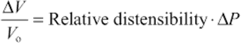
About two thirds of the total blood volume resides in the systemic veins (see pp. 448–450). When a recumbent subject assumes an upright position, the blood shifts from the central blood volume reservoirs and other veins to large veins in the dependent limbs. In discussing Figure 17-8B, we assumed that the cardiovascular system somehow made the adjustments necessary to keep right atrial pressure (RAP) constant at about +2 mm Hg. Indeed, unless compensatory mechanisms intervene, blood redistribution will lower not only arterial blood pressure but also venous return and thus cardiac output.
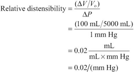
To illustrate the effect of blood redistribution on venous return, we will represent the entire circulatory system by a horizontal, distensible cylinder 180 cm in length (the height of our person) and 3 cm in radius (Fig. 25-3A). This cylinder holds ~5000 mL of blood (the normal blood volume). We know that immediately after a cardiac arrest, the entire vascular system will have a mean systemic filling pressure (MSFP) of ~7 mm Hg (see p. 549). The MSFP is the pressure in the circulation that would remain in the absence of any pumping or any gravity effects. Thus, if a subject is recumbent and has no heartbeat, and if the cardiovascular system is filled with a normal blood volume of 5000 mL (Vo), the overall compliance of the system will produce a uniform pressure of ~7 mm Hg. If we were to transfuse an additional 100 mL (ΔV) of blood into our cylinder, the MSFP would rise by ~1 mm Hg (ΔP). We can conclude that the compliance, expressed as a normalized distensibility (see p. 454), is
 (25-1)
Thus, for every 2% increase in blood volume, the MSFP of the cylinder increases by 1 mm Hg.

FIGURE 25-3 Model of the orthostatic redistribution of blood. A, A horizontal tube (3-cm radius, 180 cm long) contains the entire blood volume (5 L). With no blood flow, pressure inside the tube is uniform and corresponds to a mean systemic filling pressure of 7 mm Hg. B, With the cylinder upright (orthostasis), pressure gradually increases toward the bottom, causing increasingly greater distention of this compliant tube. Because blood volume has shifted to the bottom, the upper level of the blood column is 30 cm below the level of the heart, preventing venous return. C, Reducing compliance of the tube by half also causes distention to fall by half. With the reduced shift of blood volume, the upper level of the blood column now just reaches the heart.
What will happen to our cylinder if we now turn it upright? This position is called orthostasis (from the Greek orthos [upright] + histanai [to stand]). Because we have a vertical column of blood 180 cm tall, we must now consider gravity (see Fig. 25-3B). The highest pressures will be at the bottom of the cylinder. (Fig. 17-8 shows that orthostasis causes venous pressure at the ankle to rise from 5 to 100 mm Hg.) Therefore, our cylinder will distend maximally at the bottom, and this distention represents a shift in blood volume. The bottom of the cylinder (corresponding to the “dependent areas” of a person) gains volume, whereas the top (i.e., corresponding to the cranial portion of a person) loses blood volume. In fact, there would be no blood at all at the top of the cylinder.
By just how much would the column of blood fall in our upright, 180-cm-tall cylinder? If the overall vascular volume distensibility were 0.02/(mm Hg), then the actual height of the blood column inside the cylinder would be only about 100 cm. ![]() N25-1 If the heart were 50 cm below the top of the cylinder, then the top of the column would now be ~30 cm below the level of the heart. Therefore, there would be no blood to return to the heart. Moreover, the RAP would be negative (−30 cm H2O, or about −22 mm Hg). Because the heart cannot create a vacuum this large at its input by “sucking” blood—in fact, the heart must be filled by a positive RAP—cardiac output would fall to zero.
N25-1 If the heart were 50 cm below the top of the cylinder, then the top of the column would now be ~30 cm below the level of the heart. Therefore, there would be no blood to return to the heart. Moreover, the RAP would be negative (−30 cm H2O, or about −22 mm Hg). Because the heart cannot create a vacuum this large at its input by “sucking” blood—in fact, the heart must be filled by a positive RAP—cardiac output would fall to zero.
N25-1
Calculation of Distention in an Upright Cylinder
Contributed by Emile Boulpaep
Equation 19-4 and the identical Equation 25-1 both describe how the “relative” or “normalized” distensibility depends on the relative change in volume (ΔV/Vo) and the pressure difference (ΔP):
 (NE 25-1)
We can now solve for the relative change in volume:
 (NE 25-2)
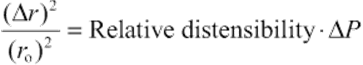
For a very thin disk of fluid (height h) in the upright vessel, the change in volume (ΔV) is due solely to a change in vessel radius. In other words, because V = hπr2, ΔV = hπ(Δr)2. Thus, we can rewrite Equation NE 25-2 as
 (NE 25-3)
In order to compute the shape of the upright vessel in Figure 25-3, the only other thing we need to know is how ΔP varies with height in the upright vessel. For a hydrostatic column, the pressure increases linearly as we descend to greater depths, as described by Equation 17-4. We reproduce this equation here:
![]() (NE 25-4)
(NE 25-4)
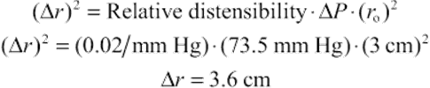
Here, ρ is the density of the liquid, g is the gravitational constant, and h is the height of the column. We could use this equation to compute the pressure in dynes per square centimeter or pascals. However, physiologists tend to express pressures in centimeters of H2O or millimeters of mercury. For example, in Figure 25-3B, the pressure at the bottom of the 100-cm column is equivalent to 100 cm H2O, which is also (knowing the density of mercury) 73.5 mm Hg.
Now we can compute the relative distention at the bottom of the upright cylinder, which is under a pressure of 73.5 mm Hg. Rearranging Equation NE 25-3 and solving for Δr (the change in radius), we have
 (NE 25-5)
Because the initial radius (ro) of the horizontal cylinder in Figure 25-3A was everywhere 3 cm, the distended radius at the bottom of the upright column has increased by 3.6 cm for a total radius of 6.6 cm—in other words, the radius has more than doubled, and the volume has more than quadrupled (i.e., increased by a factor of 4.8).
If the relative distensibility were less (e.g., 0.01/mm Hg in Fig. 25-3C), then the Δr would also be less at the bottom of the upright cylinder. On the other hand, because the Δr is less at each height, the column of water would have to be higher to accommodate the volume. Thus, the pressure at the bottom of the column would be greater (i.e., 130 cm H2O or 95.6 mm Hg in the example of Fig. 25-3C). As in Equation NE 25-5, we can compute the relative distention at the bottom of the upright cylinder, which this time is under a pressure of 95.6 mm Hg:
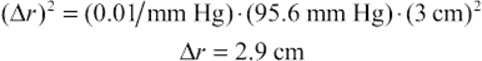 (NE 25-6)
In other words, uniformly reducing the relative distensibility by a factor of 2 still results in a near doubling of the radius and a near quadrupling of the volume (i.e., increased by a factor of 3.8).
The ANS mediates an “orthostatic response” that raises heart rate and peripheral vascular resistance and thus tends to restore mean arterial pressure
If our model predicts that orthostasis should cause RAP to fall to −22 mm Hg, how is it that the body manages to maintain RAP at about +2 mm Hg in the upright position? The answer is that pooling of blood in the dependent vessels is much less pronounced during orthostasis than would be predicted by Figure 25-3B, where ~2.3 L disappeared from the top of the cylinder. The actual amount of pooling in both legs of a real person is only ~500 mL. ![]() N25-2 Four major factors help reduce pooling and maintain RAP.
N25-2 Four major factors help reduce pooling and maintain RAP.
N25-2
Hypothetical Volume of Pooled Blood during Orthostasis
Contributed by Emile Boulpaep
In the example shown in Figure 25-3B, the column of blood would reach a height of 100 cm. Because the horizontal cylinder in Figure 25-3A had a length of 180 cm, turning the vessel upright reduced the length of the column by 80 cm. How much volume moved into the lower part of the distensible, upright vessel? The answer is the volume at the top of the white region in Figure 25-3B, which is the product of the 80-cm length and cross-sectional area:
 (NE 25-7)
Nonuniform Initial Distribution of Blood
In our cylinder example, the blood was initially distributed evenly throughout the length of the cylinder. In a recumbent human, however, most of the blood in large veins is located in the central blood volume (see p. 449), that is, the vessels near the heart. If a large fraction of the blood had started off in the head, the orthostatic shift of blood would have been more dramatic, as in Figure 25-3B. The majority of the 500 mL of blood that pools in the legs during orthostasis comes from the intrathoracic vascular compartments. What is the sequence of events by which blood volume redistributes during orthostasis? As one stands, the output from the heart for a number of beats exceeds the venous return into the thoracic pool. This excess blood ends up filling the vessels in the dependent regions of the body. The result is a net transfer of blood—by way of the heart—from the intrathoracic vascular compartments to the dependent vessels.
Nonuniform Distensibility of the Vessels
In Figure 25-3B, we assumed a relative distensibility of 0.02/(mm Hg). If we had instead used a value of 0.01/(mm Hg)—that is, if the vessels were less distensible—standing would cause a less dramatic shift of blood to the dependent vessels, ~1.4 L (see Fig. 25-3C) instead of ~2.3 L (see Fig. 25-3B). ![]() N25-1 As a result, the height of the blood column would fall from 180 to only 130 cm (see Fig. 25-3C) in the upright position, rather than to 100 cm (see Fig. 25-3B). Assuming a lower distensibility for the leg veins is reasonable because small vessels are far stiffer than larger ones, such as the aorta and vena cava. With the lower relative distensibility of 0.01/(mm Hg), the column of blood would just reach the heart. Indeed, when a subject stands quietly, the zero effective pressure level—the height in the body where vascular pressure equals atmospheric pressure—is about at the level of the right atrium. Obviously, if the circulatory system reduces its distensibility even further through the regulated contraction of vascular smooth muscle (discussed below), the height of the column of blood will increase and improve venous return.
N25-1 As a result, the height of the blood column would fall from 180 to only 130 cm (see Fig. 25-3C) in the upright position, rather than to 100 cm (see Fig. 25-3B). Assuming a lower distensibility for the leg veins is reasonable because small vessels are far stiffer than larger ones, such as the aorta and vena cava. With the lower relative distensibility of 0.01/(mm Hg), the column of blood would just reach the heart. Indeed, when a subject stands quietly, the zero effective pressure level—the height in the body where vascular pressure equals atmospheric pressure—is about at the level of the right atrium. Obviously, if the circulatory system reduces its distensibility even further through the regulated contraction of vascular smooth muscle (discussed below), the height of the column of blood will increase and improve venous return.
Muscle Pumps
An important compensation for blood pooling during orthostasis comes from skeletal muscle contraction. When a person stands, the muscles of the legs and abdomen tighten. The presence of valves in the veins, as well as intermittent muscular movement, contributes to the flow of blood upward along the veins (see Fig. 22-7C and Fig. 24-6). Vessels of the abdominal region remain nearly unaffected by orthostasis because the abdominal viscera are contained in a water-filled jacket that is maintained by the tone of the abdominal muscles.
Autonomic Reflexes
Because of decreased venous return, cardiac output tends to fall by ~20% soon after one assumes an erect position. However, the fall in cardiac output would be even greater in the absence of autonomic reflexes. The decreased venous return leads to a fall in RAP, which in turn leads to a decrease in stroke volume and thus arterial pressure. High-pressure baroreceptors (see pp. 534–535) sense this decrease in arterial pressure, ![]() N25-3 which leads to an increased sympathetic output that raises vascular tone throughout the body and increases heart rate and contractility. Together, the constriction of arterioles (which raises total peripheral resistance) and the increased heart rate restore the systemic mean arterial pressure, despite a small decrease in stroke volume. In the dependent regions of the body, the sympathetic response also increases the tone of the veins, thereby decreasing their diameter and their capacity (compare Fig. 25-3B and C).
N25-3 which leads to an increased sympathetic output that raises vascular tone throughout the body and increases heart rate and contractility. Together, the constriction of arterioles (which raises total peripheral resistance) and the increased heart rate restore the systemic mean arterial pressure, despite a small decrease in stroke volume. In the dependent regions of the body, the sympathetic response also increases the tone of the veins, thereby decreasing their diameter and their capacity (compare Fig. 25-3B and C).
N25-3
Baroreceptor Responses in Orthostasis
Contributed by Emile Boulpaep
As noted in the text (see p. 576), orthostasis leads to the following sequence of events: Decreased venous return → fall in RAP → decrease in stroke volume → decreased arterial pressure → response of high-pressure baroreceptors (see pp. 536–537) → increased sympathetic output → generalized vasoconstriction and increased heart rate/contractility. Because RAP falls early in this sequence, you might wonder what role the atrial low-pressure baroreceptors play in the response. Reduced atrial stretch has little effect on heart rate (see pp. 547–547) and causes an increase in sympathetic output only to the kidney (i.e., causing renal vasoconstriction). Therefore, the low-pressure baroreceptors make only a minor contribution to the overall orthostatic response (i.e., generalized vasoconstriction and increased heart rate/contractility).
In summary, of the four factors that contribute to the stability of RAP during orthostasis, two are anatomical (i.e., nonuniformities of initial blood volume distribution and distensibility) and two are physiological (i.e., muscle pumps and autonomic reflexes). The two physiological mechanisms are both important. Indeed, after a lumbar sympathectomy, patients tend to faint when standing. However, after some months, they are able to compensate, perhaps by using the muscle pumps more effectively or by enhancing the sympathetic response of the heart.
The extent of the orthostatic response—how much the heart rate or peripheral vascular resistance increases under the control of the ANS—depends on a variety of factors (Table 25-1), which involve nearly the entire cardiovascular system. Because these factors may differ from person to person or may differ within any one individual according to the circumstances, the orthostatic response is highly variable. We now discuss two examples of this variability.
TABLE 25-1
Factors Influencing the Degree of Orthostatic Response*
Total blood volume
Distribution of blood volume
Size of vessels in dependent regions of the body
Vascular distensibility
Mean systemic filling pressure (pressure in the absence of cardiac output)
Level at which zero effective pressure is normally located in a particular individual
Degree of tilt
Skeletal muscle tone; strength and rate of intermittent contraction of skeletal muscles
Vascular sufficiency
Abdominal muscle tone
Temperature
Response of low-pressure receptors
Response of high-pressure baroreceptors
Activity of the sympathetic system
Initial heart rate
Initial myocardial contractility
Sensitivity of vascular smooth muscle to sympathetic stimulation
*That is, by how much standing up increases heart rate and peripheral vascular resistance.
Postural Hypotension
In very sensitive subjects lying on a tilt table, a sudden orthostatic tilt can cause such a large fall in arterial pressure that the individual becomes dizzy or even faints. Fainting is caused by a transient fall in arterial pressure that causes cerebral perfusion to become inadequate.
Temperature Effects
In a cool environment, in which the arterioles in the lower extremities are constricted, the initial dip in arterial pressure can be small, despite the decrease in stroke volume. The explanation is that the high arteriolar resistance delays the transfer of blood from the thoracic pool to the legs. As a result, the sympathetic response to the small drop in mean arterial pressure may already be in effect before further pooling can occur. In a warm environment, where the arterioles in the skin are more dilated, orthostasis leads to faster transfer of blood from the thoracic pool to the legs ![]() N25-4 so that—before the sympathetic response can develop—the initial decreases in stroke volume, mean arterial pressure, and pulse pressure can be large. Thus, soldiers standing quietly at attention in hot weather are more likely to faint than are soldiers marching in a cold environment because of differences in muscle pump activity and vasoconstriction.
N25-4 so that—before the sympathetic response can develop—the initial decreases in stroke volume, mean arterial pressure, and pulse pressure can be large. Thus, soldiers standing quietly at attention in hot weather are more likely to faint than are soldiers marching in a cold environment because of differences in muscle pump activity and vasoconstriction.
N25-4
Effects of Temperature on Venous Pooling
Contributed by Emile Boulpaep
As discussed on page 565, the contraction of skeletal muscle in the legs (the “muscle pump”—see Fig. 24-6) drives blood from the large veins in the lower limbs toward the heart. Conversely, each time these skeletal muscles relax, the vascular bed in the legs refills from the arterial side. Obviously, the arteriolar inflow resistance influences the rate at which the veins in the leg refill subsequent to the action of the muscle pump. When the lower limbs are at a high temperature, the arterioles dilate, lowering the inflow resistance and increasing the inflow of blood to the dependent vessels. Therefore, venous pooling worsens at high temperature, which explains why soldiers tend to faint under these conditions.