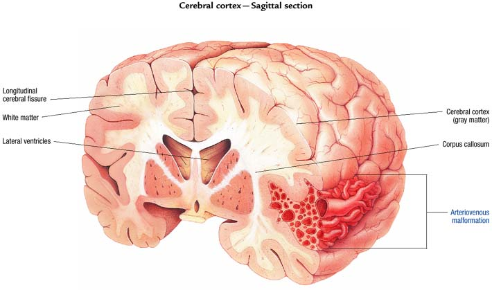
Arteriovenous malformations (AVMs) are tangled masses of thin-walled, dilated blood vessels between arteries and veins that aren't connected by capillaries. AVMs are common in the brain, primarily in the posterior portion of the cerebral hemispheres. Abnormal channels between the arterial and venous system mix oxygenated and unoxygenated blood and thereby prevent adequate perfusion of brain tissue.
AVMs range in size from a few millimeters to large malformations extending from the cerebral cortex to the ventricles. Usually, more than one AVM is present. Males and females are affected equally, and some evidence exists that AVMs occur in families.
![]() Age Alert
Age Alert
Most AVMs are present at birth; however, symptoms may occur at any time. Two-thirds of all cases occur before age 40.
Causes
· Congenital: hereditary defect
· Acquired: trauma such as penetrating injuries
Pathophysiology
AVMs lack the typical structural characteristics of the blood vessels. The vessel walls of an AVM are very thin; one or more arteries feed into the AVM, causing it to appear dilated and torturous. The typically high-pressured arterial flow moves into the venous system through the connecting channels to increase venous pressure, engorging and dilating the venous structures. An aneurysm may develop. If the AVM is large enough, the shunting can deprive the surrounding tissue of adequate blood flow. Additionally, the thin-walled vessels may ooze small amounts of blood or actually rupture, causing hemorrhage into the brain or subarachnoid space.
Signs and symptoms
Typically, few or none.
If AVM is large, leaks, or ruptures
· Chronic headache and confusion
· Seizures
· Systolic bruit over carotid artery, mastoid process, or orbit
· Focal neurologic deficits (depending on the location of the AVM)
· Hydrocephalus
· Paralysis
· Loss of speech, memory, or vision
![]() Clinical Tip
Clinical Tip
Symptoms of intracranial hemorrhage, indicating AVM rupture, include sudden severe headache, seizures, confusion, lethargy, and meningeal irritation.
Diagnostic test results
· Cerebral arteriogram confirms the presence of AVMs and evaluates blood flow.
· Doppler ultrasonography of the cerebrovascular system indicates abnormal, turbulent blood flow.
Treatment
· Supportive measures, including aneurysm precautions
· Surgery, including block dissection, laser, or ligation
· Embolization or radiation therapy
· Radiosurgery
P.125
ARTERIOVENOUS MALFORMATION
|
|
|
|