George Iancu1
(1)
Department of Obstetrics and Gynecology, “Carol Davila” University of Medicine and Pharmacy Filantropia Clinical Hospital, 11-13, Blvd. Ion Mihalache, Bucharest, 71117, Romania
George Iancu
Email: klee_ro@yahoo.com
Abstract
Obstetric fistula is common in developing countries as a result of unattended prolonged obstructed labour. Evaluation of obstetric fistula is based on history and clinical examination. Continuous urinary or stool leakage started soon after a long labour that ended usually with stillbirth in a low resource area is highly suggestive of obstetric fistula. To date, there is no consensus regarding the classification of obstetric fistula. Multiple classification systems have been proposed, but all the present classifications of obstetric fistula are of limited clinical use because of the lack of impact on treatment outcome. Surgical treatment with closure of fistulous tract and reconstruction of local anatomy is the mainstay of therapy. Persistent urinary incontinence after successful surgical repair is the most concerning problem; additional continence surgery is usually needed. In experienced hands, the success rate of surgery varies around 80–90 % after the first surgical repair. Postoperative care should be focused on appropriate bladder drainage and early identification of complications.
Keywords
Obstetric fistulaObstructed labourUrinary incontinenceLow-resource areaIncontinence surgeryFistula recurrenceContinuous bladder drainage
Background and Definition
The genital fistula is an abnormal communication between the genital tract and urinary or digestive system. Fistula is classified as genitourinary or genito-anorectal fistula depending on the type of communication. In developed countries, most fistulae are the consequence of surgery, while in developing countries, it is the obstetric cause that leads. The maternal mortality and morbidity due to inappropriate access to medical resources is still unacceptably high in developing countries [1, 2]. Obstetric fistula is one of the most difficult to manage complications of a dystocic delivery with a great impact on women’s quality of life.
Epidemiology and Risk Factors
The incidence of obstetric fistula cited by World Health Organization is about 0.3 % of deliveries. The true prevalence of the disease is unknown. There are epidemiological studies that estimate the incidence of obstetric fistula to be around 0.35 % of deliveries [3]. The prevalence is reported to be high especially in Africa, in countries such as Sudan, Ethiopia, Ghana, Nigeria and in South Asia, mainly Bangladesh [4]. The prevalence of the condition is likely to be even higher in the rural areas of the developing world where access to healthcare facilities is usually difficult and available data are limited.
Risk factors associated with obstetric fistula are primiparity, young age, delivery of a large fetus, malpresentation, short statured patient, lack education [5, 6].
Pathogenesis
The aetiology of obstetric fistula is mainly ischaemic due to prolonged obstructed labour. The main cause is probably cephalopelvic disproportion, fistulae being often located at the bladder neck or vault. The mother’s soft tissues are compressed against the bony pelvis during labour that lasts usually for several days. The continuous pressure diminishes the blood supply to the soft tissues and generates extensive damage with tissue necrosis and fistula, scarring and alteration of normal pelvic anatomy. Often, the injury involves not only the bladder and the vagina, but the urethra, uterus or rectum as well; urogenital, gastrointestinal, neurologic and musculoskeletal systems are usually involved. Other causes of obstetric fistula encountered less frequently are iatrogenic trauma during caesarean section, difficult instrumental deliveries or other obstetric maneuvers [7].
Clinical Diagnosis
Symptoms and Signs
The obstetric fistula is usually a consequence of prolonged abnormal labour; one study estimated the average length of labour of almost 4 days (mean 3.9 days, range 1–6), with 92.7 % ending as stillbirths [5]. Usually, it takes about 3–10 days for the communication to develop after the necrotic ischaemic slough is drained out through the vagina (Fig. 15.1). The patient becomes continuously incontinent after fistula formation. Some patients are rendered incontinent for stool, if a rectovaginal fistula has concomitantly developed. Obstructed labour trauma that results in fistula formation is different from the surgical injury; it involves often other organ systems as well and the fistula is usually wider due to extensive ischaemic injury. The average size of obstetric fistula treated in a fistula hospital in Addis Ababa was 2.3 cm long (range 0.1–8 cm) and 2.5 cm wide (range 0.1–10 cm) [8].
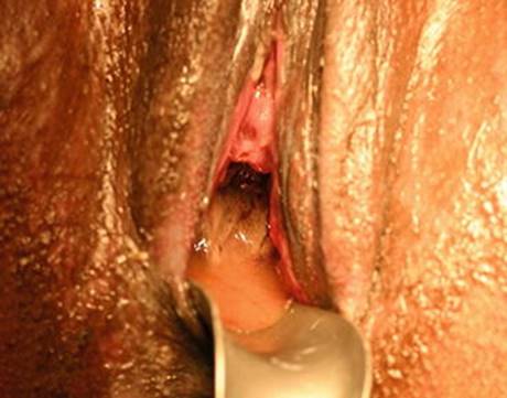
Fig. 15.1
Early vesicovaginal fistula with sloughing tissue draining out through the vagina (Reproduced with permission of Dr. Andrew Browning)
Vesicovaginal fistulae are the most common type of fistulae. The site of vesicovaginal fistula varies depending on the level of impaction of labour; if the mechanical conflict is at the level of the pelvic inlet, usually the fistula develops intra- or juxtacervical (Fig. 15.2). If the impaction occurs lower in the pelvis, usually the urethra is involved (Fig. 15.3), with severe compromise of the continence mechanisms in the long term [9]. Urethral involvement is usually a predictor for poor prognosis regarding the continence outcome [10]. It can occur in up to one third of obstetric fistula patients and about 5 % of the cases can present total urethral loss [8]. Ureteral lesions can lead to genito-urinary fistula as well; in a small number of cases, involvement of the distal ureter is followed by uretero-vaginal fistula, with continuous free drainage of urine into the vagina [11]. The fistulous tract can involve the uterus as well, though more rare and usually due to operative injury after caesarean section [12]. Usually, they manifest as vaginal urinary leakage or sometimes as cyclical haematuria.
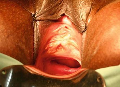
Fig. 15.2
Large vesicovaginal fistula at the level of midvagina, juxtacervical (Reproduced with permission of Dr. Andrew Browning)
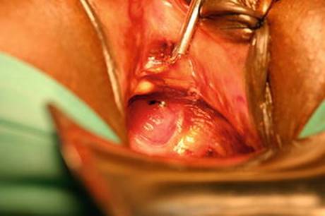
Fig. 15.3
Small circumferential urethrovaginal fistula (Reproduced with permission of Dr. Andrew Browning)
Obstetric trauma can involve the digestive tract as well. Most commonly, rectovaginal fistulae are the consequence of fetal impaction against the rectum, followed by ischaemic necrosis of the rectovaginal septum. A study revealed a prevalence of rectovaginal fistula of 1–8 % of obstetric fistula and 1–23 % for combined vesico-vaginal and recto-vaginal fistula; vesico-vaginal fistula accounted for the vast majority of obstetric fistula (over 80 %) [13]. The level of the fistula is important because involvement of anal sphincter can compromise the fecal or flatal continence mechanism.
In developing countries, genitourinary fistulae can be associated with upper renal tract damage, from mild hydronephrosis to non-functioning kidney requiring nephrectomy [11, 14]. The upper urinary tract damage is usually secondary to obstructive uropathy caused by the scarred ureter. Bladder stones form due to recurrent infections, reduced water intake followed by concentrated urine, insertion of foreign bodies in the vagina that act as promoters for calculogenesis [7]. The continuous leakage of urine irritates the perineal skin, causing dermatitis, excoriations, superficial infections or hyperkeratosis.
Regarding the extent of the changes of the reproductive tract, these vary from minimal or mild changes in anatomy (though this is usually the case with surgical fistula) to extensive damage, with vaginal injuries up to necrosis of the whole vagina, torn cervix and involvement of the uterus. Vaginoplasty is required in about one third of the cases [8].
The reproductive outcome is frequently severely affected. Amenorrhoea occurs in half of the patients with obstetric fistula [8]. The aetiology has been debated; amenorrhoea is probably due to the stress of delivery and presence of fistula leading to social isolation, low BMI, pituitary failure after obstetric haemorrhage or shock in long labour, Asherman’s syndrome or obstructed outflow and subsequent haematometra [7]. The pregnancy rate in a patient previously treated for fistula is as low as 19 % [15]; small series of pregnant patients delivering post fistula repair showed high recurrence rates of fistulae after vaginal delivery (27 %) and good outcomes with no recurrence after caesarean delivery [16, 17].
Apart from the local anatomical changes related to the pathophysiology of the disease, there are also associated conditions that manifest commonly in patients with obstetric fistula. Obstetric fistula is usually associated with social consequences and an important impact on mental health due to its circumstances of occurrence: young women from low resource country that laboured for days and usually lost the baby, divorced and living in isolation because of the debilitating condition [5, 18]. The vast majority of fistula patients present with mental health problems; one study revealed 97 % of the patients with obstetric fistulae screened positive for mental disorders [19]. Other associated conditions cited in the literature are malnutrition or limb contractures [7].
Assessment
History and clinical examination are the first tools for the assessment of a patient with suspected obstetric fistula. Continuous incontinence that started soon after a long labour, ending usually with stillbirth, in a low resource area is characteristic for obstetric fistula. The clinical examination will enable the diagnosis of a large fistula, its location and the extent of scarring. If the size of the fistula is small and it cannot be identified during naked eye examination, a dye test can be undertaken. Swabs are placed in the vagina, the bladder is catheterized and methylene blue is retrograde injected in the bladder. After a few minutes, the swabs are checked for leakage. After identifying and localizing the genitourinary fistula, the posterior vaginal wall is carefully checked for rectovaginal fistula. The integrity of anal sphincter should be ascertained as well. When a rectovaginal fistula is identified close to the external anal sphincter, reconstruction of the sphincter might be required. Sometimes, when the patient is symptomatic (flatus or stool incontinence) and there is no obvious fistula on examination, a small fistula can be diagnosed using instillation of dye per rectum with the aid of a Foley catheter.
Other more complex diagnostic tests (intravenous urography etc) may be required to establish a diagnosis of fistula. The fistula location and its relationship with the ureteral orifices and urethra are very important to plan management. Large fistulae may allow the identification and catheterization of ureteral orifices vaginally through the fistula tract. Cystoscopy is helpful for small fistulae or for fistulae located high, and are difficult to be accessed vaginally.
Classification of Fistulae
To date, there is no consensus regarding the classification of obstetric fistulae. A valid classification should follow criteria according to the impact on treatment outcome; there are no prospective studies to evaluate the prognosis of different categories of fistulae. Accordingly, all the present classifications of obstetric fistulae are of limited clinical use concerning the impact on treatment outcome [20].
The World Health Organization (WHO) proposed a classification of obstetric fistulae. This classification takes into account the difficulty of the surgical repair. Depending on the complexity, there are two types of fistulae [21]:
· Simple fistulae, with good prognosis, that can be repaired by surgeons trained to treat uncomplicated fistula; they are usually single vesico-vaginal fistulae, under 4 cm diameter, without urethral or ureteral involvement, minimal vaginal scarring and tissue loss, no previous fistula surgery (Fig. 15.4).
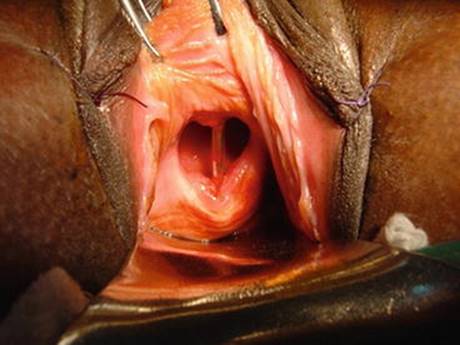
Fig. 15.4
Medium vesicovaginal fistula at the level of midvagina. No urethral involvement is seen (Reproduced with permission of Dr. Andrew Browning)
· Complicated fistula, requiring usually referral to be repaired only by specialist fistulae surgeons; they are multiple, recto-vaginal or combined vesico- and recto-vaginal, involvement of cervix, urethra, ureters draining into the vagina, vaginal tissue scarring, circumferential loss, recurrent fistulae after failed repairs (Fig. 15.5).
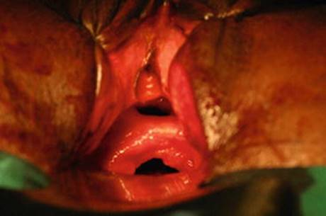
Fig. 15.5
Complicated fistula: double urethrovaginal and vesicovaginal, circumferential, with almost complete urethral loss (Reproduced with permission of Dr. Andrew Browning)
Other classification systems have also been proposed (Table 15.1).
Table 15.1
Classification systems of vesicovaginal fistulae of obstetric origin
|
Author (year) |
Criteria considered |
Classification/type |
|
Waaldijk (1995) [22] |
Urethral closing mechanism involvement |
I. Urethral closing mechanism intact IIAa. Urethal closing mechanism affected, without (sub)total urethral involvement or circumferential defect IIAb. Without (sub)total urethral closing mechanism involvement, with circumferential defect IIBa. (Sub)total urethral involvement, without circumferential defect IIBb. (Sub)total urethral involvement and circumferential defect III. Ureteric involvement; other rare fistulae. |
|
Browning (2004) [9] |
Vaginal scarring Bladder volume |
1. Simple – reduced vaginal scarring and normal bladder volume 2. Complex – severe vaginal scarring and/or reduced bladder volume; needs vaginoplasty or reconstruction |
|
Goh (2004) [23] |
Distance to urethral meatus Size of fistula Vaginal fibrosis/length/capacity |
1. distal edge > 3.5 cm from external urethral meatus (EUM) 2. distal edge at 2.5–3.5 cm from EUM 3. distal edge 1.5–2.5 cm from EUM 4. distal edge <1.5 cm from EUM (a) size < 1.5 cm in maximal diameter (b) size 1.5–3 cm in maximal diameter (c) size > 3 cm in maximal diameter (i) minimal or absent vaginal/perifistular fibrosis, vaginal length > 6 cm, normal vaginal capacity (ii) moderate to severe fibrosis and/or reduced vaginal length and/or capacity (iii) special considerations (ureteric involvement, previous repair, circumferential fistula) |
The prognostic factors associated with the outcome of the repair of obstetric fistula are the size of the fistula, concurrent lesions (rectovaginal fistula), degree of scarring around the fistula and involvement of the continence mechanism, including urethral damage [10, 24]. Apart from the factors already mentioned, the vaginal route of repair instead of abdominal route and duration of catheterization more than 14 days have been associated with increased risk of failure of the repair [25].
Management
When the management of obstetric fistula is considered, general aspects should be discussed (Table 15.2).
Table 15.2
General considerations in obstetric fistula management
|
Aspects of management of obstetric fistula |
|
Timing of surgery |
|
Abdominal/vaginal approach |
|
Excision/conservation of fistulous tract |
|
Tissue interposition: |
|
Omental flap |
|
Labial fat pad (Martius graft) |
|
Peritoneal flap |
|
Muscle flap |
|
Concomitant procedures: stress urinary incontinence treatment, cystoplasty, vaginoplasty |
|
Adjuvant treatment: postoperative drainage, anticholinergic therapy, antibiotics, HRT |
The timing of repair is very important especially for obstetric fistula where the main aetiologic factor is extensive ischaemic necrosis; the time of surgical intervention should be carefully selected, due to the need for good quality tissue for the fistula to heal properly and to avoid recurrences. Usually, it is recommended to wait for at least 3–6 months from the causative injury. The waiting time should allow the necrotic tissue to separate from the normal one that will be used in the repair. Some authors advocate for immediate repair to avoid issues related to potential social rejection experienced by the patient. They claim, there is no significant difference between early repair and the classic repair performed after a few months. The cited study consisted of more than 1700 cases operated by an experienced fistula surgeon with roughly 95 % success rate for first attempt repair; however, the results might not be reproducible in other services with less experience in fistula treatment [26].
Early treatment of fistula could be achieved using continuous bladder drainage as sole therapy, especially for a small fistula that can be closed conservatively thus avoiding surgery. The bladder in this circumstance should be kept on free drainage for 3–4 weeks depending on the fistula size and extension of necrosis. The reported cure rates vary between 7 and 15 % [26, 27]. The success rates depend on the degree of atrophy secondary to menopause, size of the fistula or scarring extent. If closure does not occur after 4 weeks of continuous drainage, surgical treatment is usually needed [28].
A fistula can be repaired vaginally or abdominally. The route of repair depends on the accessibility to the surgical site and experience and skills of the surgeon. The abdominal route is used for vault fistula, juxtacervical or vesicouterine location [7]; ureteral injury, need for augmentation cystoplasty or concomitant abdominal pathology mandate abdominal approach as well. The abdominal approach is associated with morbidity related to laparotomy and requires cystotomy to access fistula site. When the exposure of the superior vagina is difficult, relaxing perineal incisions can be used to facilitate accessibility via the vaginal route (Fig. 15.6) [29]. A study suggested though that for particular indications, the abdominal approach might have better success rate than the vaginal route. Factors like extensive scarring, ureteric, trigonal or supratrigonal involvement, vesicouterine or vesicocervical location were followed by better outcomes of the repair when approached transabdominally; the relative risk of failure for vaginal approach was 1.41 [30].
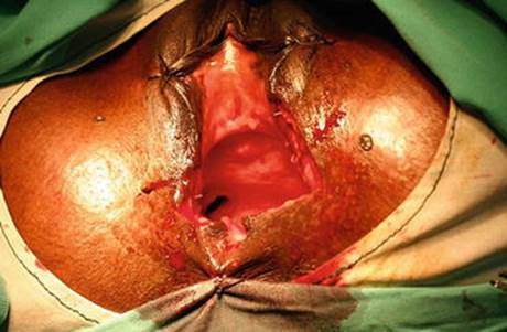
Fig. 15.6
Relaxing perineal incision for juxtacervical ureterovaginal fistula with narrowed introitus (Reproduced with permission of Dr. Andrew Browning)
Regarding the excision of fistula tract, the opinions are divided. Some authors suggest excising it to reduce the recurrence rate, while others advocate a surgical repair without excising the fistula tract which would increase the fistula size and might require electrocautery to control bleeding, thus creating more nonviable tissue. The fistula tissue provides good support for the first layer of sutures. It is important to dissect and mobilize the bladder wall in order to avoid any tension on the repair site with suturing. Care should be taken to avoid ureteral injuries, during surgical repair. The ureters should be catheterized for all fistulae involving the trigone or located supratrigonally [7]. Because of the big size of obstetric fistulae, ureteral catheterization can often be undertaken through the fistula tract.
A flap of tissue or graft is occasionally used to repair a fistula involving urethra or urethrovesical junction to limit the scarring and avoid recurrence. The options are labial or bulbospongiosus fat pad (Martius graft), omental flap, peritoneal flap, gluteal or gracilis flap [29]. The use of tissue interposition techniques optimized the success rate of the procedure in a couple of studies [31, 32]. However, the use of vascular tissue flaps is still controversial. Authors with vast experience in fistula surgery report similar success rates independent of the graft use [33].
Most commonly, the graft used is harvested from the labia majora (Martius graft); a longitudinal incision is made along the labia majora, exposing the underlying fat. The flap of fat is then developed, conserving the posterior vascular pedicle to ensure viability. The flap is then rotated medially, reaching the fistula site through a tunnel created in front of the pubic ramus and behind the bulbospongiosus muscle. The flap is anchored at the site of fistula to promote healing. Some surgeons drain the labial site to prevent hematoma formation.
Important principles that should be followed for the surgical treatment of fistula include mobilization of the fistulous tract enough to enable closure without tension, water-tight closure of the injury site and careful postoperative management, providing adequate bladder drainage to promote wound healing [34].
When approaching the fistula vaginally, the patient is placed in lithotomy position and the entire fistula tract is exposed. The vaginal opening of the fistula is incised circumferentially and the fistula mobilized so that the fistula margins can be brought together without tension to close the communication between urinary and genital tract. After mobilization of the fistulous tract and the surrounding tissue, the edges of the fistula are closed with absorbable material, either interrupted or continuous depending on the surgeon’s preference. The second layer of sutures in the bladder wall could be considered in the absence of extensive scarring. It is very important to ensure that the suture in the bladder wall is water-tight. Therefore, 100–250 ml of diluted methylene blue or indigo carmine dye are instilled in the bladder and the suture line is checked for leakage. If leakage occurs, revision of the suture line is considered until it is water-tight. The instilled fluid should not exceed 250 ml to avoid bladder over-distension and break-down of the fresh repair. The vaginal epithelium is closed with running absorbable sutures.
Urinary Incontinence After Repair
Persistent urine loss after fistula repair can have multiple causes. Recurrence of fistula should be excluded first: the patient should be examined to rule out a recurrence and if transurethral incontinence is diagnosed, other causes of incontinence should be considered. Frequently, urinary incontinence occurs after surgical treatment of fistula, either due to loss of the sphincteric mechanism of vesicourethral junction and urethral injury or reduced bladder capacity and urgency urinary incontinence. Browning assessed the risk of urinary incontinence after fistula closure on 318 patients and divided them into two groups: simple fistulae with minimal scarring and good bladder volume and complex fistulae with severe scarring and/or reduced bladder volume. The risk of incontinence after fistula repair was 50 % in the first group and 100 % in the complex fistulae group [9].
Persistent fistula is ruled out by vaginal examination and dye test. If leakage through the repair site is diagnosed, it is advisable to wait for 6–8 weeks to allow healing and thereafter to consider another surgical intervention if the leakage persists. If the fistula tract is small, continuous bladder drainage can result in spontaneous closure.
When transurethral incontinence is diagnosed and a recurrent fistula is excluded, the type of urinary incontinence should be ascertained. Urodynamic tests are mandatory for the evaluation of these patients; unfortunately, in resource-limited settings, urodynamic facilities are usually not available. A recent study on urodynamic evaluation of residual urinary incontinence after obstetric fistula repair showed a prevalence of almost 50 % for stress incontinence, over 40 % mixed urinary incontinence and only 3 % detrusor overactivity [35].
The management of patients treated for urogenital fistula and diagnosed with postoperative transurethral urinary incontinence proves to be often difficult especially in cases of preexisting damage of the urethral sphincter, reduced bladder capacity and detrusor overactivity. The use of synthetic mesh sling in patients with a history of obstetric fistula was associated with higher rates of erosion and lower rates of success, advocating the use of autologous sling in this clinical scenario [36]. The use of a urethral plug was proposed as a non-invasive therapy for stress urinary incontinence after fistula repair. The plug should be removed every three hours by the patient to allow bladder emptying [37].
Urinary diversion and bladder augmentation are therapeutic measures of last resort for patients with persistent urinary incontinence [34]. They are sometimes the only options for patients with most of the bladder injured. Patients with bladder augmentation with intact urethra often require intermittent self-catheterization that may not be an option in low resource areas due to lack of catheters and equipment. Urinary diversion can be achieved by ureterosigmoidostomy, Mainz II pouch or ileal conduit [7].
Particular Clinical Forms of Urogenital Fistulae/Complex Fistulae
The criteria to consider a fistula complex are variable. The size of the fistula is important (greater than 4–6 cm), involvement of the continence mechanism (urethra partially or completely absent, bladder capacity reduced), recurrent fistulae, extensive scarring or association of rectovaginal fistula.
Circumferential Fistula
This type of fistula remains one of the most challenging to repair. It is a result of complete transection of the urethra that is separated from the bladder. It is usually due to impaction of the presenting part against the symphysis pubis. In the vast majority of patients, the continence mechanism is impaired and needs to be addressed usually at the time of the repair. The surgical correction requires extensive mobilization of the bladder from the vagina and symphysis pubis and re-anastomosis to the urethra.
Complete/Partial Absent Urethra
Extensive damage and large defects are common features of obstetric fistula. When obstructed labour injury involves the urethra causing it to slough away (Fig. 15.7), surgical reconstruction that restores the anatomy and continence is challenging for the surgeon. This clinical form was recognized decades ago and its management was largely debated; a cohort of 50 women with totally destroyed urethra after prolonged obstructed labour was published in 1969. The urethra was reconstructed using skin and connective tissue covering the pubic bone and inferior margin of the symphysis pubis. The reconstructed urethra was reinforced with gracilis muscle and/or labial fat graft [38].
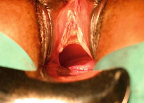
Fig. 15.7
Large circumferential vesicovaginal fistula at the level of midvagina with partial absent urethra (Reproduced with permission of Dr. Andrew Browning)
Extensive Vaginal Injury
For large fistulae with extensive scarring and damage of the vaginal wall, vaginoplasty is usually recommended. It can be required in about one third of the obstetric fistula cases [16]. Depending on the extent of vaginal scarring, reconstructive procedures can vary from a Fenton type procedure to complex vaginal reconstruction with rotational flaps use from labia or gluteal skin or other tissue (sigmoid, ileum [39]).
Rectovaginal Fistulae
The coexistence of rectovaginal fistula with urogenital fistula increases the complexity of therapeutic management. The association between the two was estimated in one large cohort to be 17 %; rectovaginal fistulae alone were found in 4 % of patients [40]. The principles of management are essentially the same: mobilization of fistula, excision of excessive scar tissue, closure of the fistula tension free, use of grafts (Martius) rarely necessary, closure of vaginal epithelium. The surgical repair of rectovaginal fistula has a lower success rate than for vesicovaginal fistula, regardless of the association of the two, but the continence outcome after successful repair of rectovaginal fistula is better than after vesicovaginal fistula [8].
Outcomes and Complications of Obstetric Fistula Surgery
The success rates after fistula surgery vary because of different definitions used by different authors. Success of fistula repair is commonly reported either as closure of abnormal communication or defect or as continence after repair or dryness. Currently, there is a lack of consensus regarding definitions and clinical success related to obstetric fistula [41]. Usually, success is considered the closure of the fistula without considering the continence outcome or patient’s quality of life. The reported success rate for fistula of obstetric origin varies among different authors, depending on the surgeon’s expertise, severity and complexity of fistula and possibly surgical technique used (Table 15.3). Success rates vary essentially between 80 and 90 % in the majority of studies; success rates over 95 % are usually reported by surgeons with vast experience in fistula treatment, working in centers with great workload. Particularities of fistula such as degree of scarring, urethral involvement, fistula size and location, circumferential fistula or reduced bladder capacity, history of failed repair can be used to stratify the prognosis. Other factors, such as female genital mutilation, parity or antibiotic use are not predictors for fistula outcome [10, 42].
Table 15.3
Success rates after obstetric fistula surgery
|
Author |
No of patients |
Success rate |
|
Rafique et al. (2002–2003) [45] |
42 patients |
85.7 % |
|
Husain et al. (2005) [46] |
50 patients |
63 % after primary repair 61 % after multiple repairs |
|
Singh et al. (2010) [27] |
42 patients |
80.1 % after primary repair |
|
Arrowsmith et al. (1994) [47] |
98 patients |
81 % after primary repair 96 % after multiple repairs |
|
Kliment et al. (1992) [48] |
41 patients |
85.4 % after primary repair |
|
Kayondo et al. (2011) [49] |
77 patients |
77.9 % after primary repair |
|
Chigbu et al. (2006) [50] |
78 patients |
82 % after primary repair |
|
Morhason-Bello et al. (2008) [51] |
71 patients |
79.2 % after primary repair |
|
Rijken et al. (2007) [44] |
407 patients |
94.1 % after primary repair |
|
Hilton (2012) [52] |
348 pt (2/3 gynecologic origin, 1/3 obstetric origin) |
95.7 % overall (98.2 % – primary repair; 88.2 % – previously failed repairs) |
|
Browning et al. (2006) [33] |
413 patients |
97.6 % after primary repair |
|
Browning et al. (2007) [53] |
316 patients |
97.5 % after primary repair |
|
Nardos et al. (2012) [54] |
189 patients |
95.2 % after primary repair |
|
Waaldijk K (2004) [26] |
1716 patients |
95.2 % after primary repair |
Regarding the time frame when success should be assessed, there are different opinions as well. Browning et al. (2008) suggest that persistent incontinence after fistula repair improves usually after 6 months [43]. Rijken et al. (2007) reported that over 50 % of cases of persistent incontinence after repair resolved by 6 months follow-up [44].
Apart from persistent urinary incontinence after surgical repair, which is the most concerning complication and has already been discussed, there are complications such as voiding difficulty and incomplete bladder emptying; they are consequence of neuropathic injury due to obstructed labour trauma and extensive dissection to mobilize a fistula located at the trigone. The rate of erosion associated with synthetic suburethral sling in obstetric fistula patients is higher than in non-fistula patients [36]. Biological slings are often preferred for urinary incontinence treatment in patients with history of fistula [9, 55, 56]. Bladder stones develop when the repair was performed using non-absorbable sutures. Other complications are common to other types of gynecologic surgery as well (Table 15.4).
Table 15.4
Complications after fistula surgery
|
Recurrent fistula |
|
Urinary/fecal incontinence |
|
Ureteral injury |
|
Reduced bladder capacity |
|
Urinary tract infection |
|
Voiding dysfunction (urinary retention to anuria) |
|
Bladder stones |
|
Amenorrhea |
|
Vaginal stenosis/atresia |
|
Asherman’s syndrome |
|
Leg weakness, contractures |
|
Haemorrhagic complications |
|
Wound infection |
Postoperative Care
Careful continuous drainage of the bladder for 10–14 days postoperatively with nursing support is the mainstay of the postoperative management for patients with urogenital fistula [54]. A full bladder could result in pressure on the repair site and failure of the surgical treatment. Imaging studies (cystogram, CT scan) may be indicated to be performed prior to the removal of catheter [57]; this can be problematic in low-resource settings. Ureteral catheters should be removed at the end of the procedure if ureteral orifices are far from the repair site or kept in situ for 5–7 days if they are in its proximity. Vaginal packing for 24–48 hours should be placed to ensure compressive haemostasis. Early mobilization and high fluid intake are also advisable.
Obstetric fistula continues to represent an important burden for the women’s health internationally. Occurring mainly in developing countries, it urges mobilization of medical resources to improve access to healthcare facilities and provide adequate maternity care.
References
1.
Conde-Agudelo A, Belizan JM, Lammers C. Maternal-perinatal morbidity and mortality associated with adolescent pregnancy in Latin America: cross-sectional study. Am J Obstet Gynecol. 2004;192:342–9.CrossRef
2.
Patton GC, Coffey C, Sawyer SM, Viner RM, Haller DM, Bose K, et al. Global patterns of mortality in young people: a systematic analysis of population health data. Lancet. 2009;374:881–92.CrossRefPubMed
3.
Harrison KA. Child-bearing, health and social priorities: a survey of 22, 774 consecutive hospital births in Zaria, Northern Nigeria. Br J Obstet Gynaecol. 1985;92 Suppl 5:1–119.PubMed
4.
United Nations Population Fund and Engender Health. Obstetric fistula needs assessment report: findings from nine African countries. New York: United Nations Population Fund and Engender Health; 2003.
5.
Kelly J, Kwast BE. Epidemiological study of vesico-vaginal fistulas in Ethiopia. Int Urol J. 1993;4:278–81.
6.
Wall LL, Karshima JA, Kirschner C, Arrowsmith SD. The obstetric vesicovaginal fistula: characteristics of 899 patients from Jos, Nigeria. Am J Obstet Gynecol. 2004;190:1011–9.CrossRefPubMed
7.
Browning A. Urogenital fistulae – obstetric, Chapter 89. In: Cardozo L, Staskin D, editors. Textbook of female urology and urogynecology, vol. 2. Boca Raton: Informa Healthcare; 2006.
8.
Arrowsmith S, Hamlin EC, Wall LL. Obstructed labor injury complex: obstetric fistula formation and the multifaceted morbidity of maternal birth trauma in the developing world. Obstet Gynecol Surv. 1996;51(9):568–74.CrossRefPubMed
9.
Browning A. Prevention of residual urinary incontinence following successful repair of obstetric vesico-vaginal fistula using a fibro-muscular sling. BJOG. 2004;111(4):357–61.CrossRefPubMed
10.
Frajzyngier V, Ruminjo J, Barone MA. Factors influencing urinary fistula repair outcomes in developing countries: a systematic review. Am J Obstet Gynecol. 2012;207(4):248–58. doi:10.1016/j.ajog.2012.02.006. Epub 2012 Feb 20.CrossRefPubMedPubMedCentral
11.
Benchekroun A, Lachkar A, Soumana A, Farih MH, Belahnech Z, Marzouk M, Faik M. Uretero-vaginal fistulas. 45 cases. Ann Urol (Paris). 1998;32(5):295–9.
12.
Tazi K, el Fassi J, Karmouni T, Koutani A, Ibn Attya AI, Hachimi M, et al. Vesico-uterine fistula. Report of 10 cases. Prog Urol. 2000;10(6):1173–6.PubMed
13.
Tebeu PM, Fomulu JN, Khaddaj S, de Bernis L, Delvaux T, Rochat CH. Risk factors for obstetric fistula: a clinical review. Int Urogynecol J. 2012;23(4):387–94. doi:10.1007/s00192-011-1622-x. Epub 2011 Dec 6.CrossRefPubMed
14.
Lagundoye SB, Bell D, Gill G, Ogunbode O. Urinary tract changes in obstetric vesico-vaginal fistulae: a report of 216 cases studied by intravenous urography. Clin Radiol. 1976;27(4):531–9.CrossRefPubMed
15.
Aimaku VE. Reproductive functions after the repair of obstetric vesicovaginal fistulae. Fertil Steril. 1974;25:586–91.CrossRef
16.
Evoh NJ, Akinla O. Reproductive performance after the repair of obstetric vesico-vaginal fistulae. Ann Clin Res. 1978;10(6):303–6.PubMed
17.
Browning A. Pregnancy following obstetric fistula repair, the management of delivery. BJOG. 2009;116(9):1265–7. doi:10.1111/j.1471-0528.2009.02182.x. Epub 2009 May 11.CrossRefPubMed
18.
Mselle LT, Moland KM, Evjen-Olsen B, Mvungi A, Kohi TW. “I am nothing”: experiences of loss among women suffering from severe birth injuries in Tanzania. BMC Womens Health. 2011;11:49. doi:10.1186/1472-6874-11-49.CrossRefPubMedPubMedCentral
19.
Goh JT, Sloane KM, Krause HG, Browning A, Akhter S. Mental health screening in women with genital tract fistulae. BJOG. 2005;112(9):1328–30.CrossRefPubMed
20.
Wall LL. Obstetric vesicovaginal fistula as an international public-health problem. Lancet. 2006;368:1201–9.CrossRefPubMed
21.
WHO. Obstetric fistula – Guiding principles for clinical management and programme development –Department of Making Pregnancy Safer; 2006.
22.
Waaldijk K. Surgical classification of obstetric fistula. Int J Gynecol Obstet. 1995;49:161–3.CrossRef
23.
Goh JT. A new classification for female genital tract fistula. Aust N Z J Obstet Gynecol. 2004;44(6):502–4.CrossRef
24.
Wall LL, Arrowsmith SD, Briggs ND, Browning A, Lassey AT. The obstetric vesicovaginal fistula in the developing world. Obstet Gynecol Survey. 2005;60 Suppl 1:S1–51.
25.
Frajzyngier V. Toward a better understanding of urinary fistula repair prognosis: results from a multi-country prospective cohort study. Columbia University, Department of Epidemiology: Dissertation; 2011.
26.
Waaldijk K. The immediate management of fresh obstetric fistulas. Am J Obstet Gynecol. 2004;191(3):795–9.CrossRefPubMed
27.
Singh O, Gupta SS, Mathur RK. Urogenital fistulas in women: 5-year experience at a single center. Urol J. 2010;7(1):35–9.PubMed
28.
Miller EA, Webster GD. Current management of vesicovaginal fistulae. Curr Opin Urol. 2001;11(4):417–21.CrossRefPubMed
29.
Schlunt Eilber K, Rosenblum N, Rodriguez L. Chapter 53 – Vesicovaginal Fistula: Complex Fistulae. In: Vasavada SP, editor. Female urology, urogynecology and voiding dysfunction. New York: Marcel Dekker; 2005. p. 761–82.
30.
Frajzyngier V, Ruminjo J, Asiimwe F, Barry TH, Bello A, Danladi D, et al. Factors influencing choice of surgical route of repair of genitourinary fistula, and the influence of route of repair on surgical outcomes: findings from a prospective cohort study. BJOG. 2012;119(11):1344–53.CrossRefPubMedPubMedCentral
31.
Evans DH, Madjar S, Politano VA, Bejany DE, Lynne CM, Gousse AE. Interposition flaps in transabdominal vesicovaginal fistula repairs: are they really necessary? Urology. 2001;57:670–4.CrossRefPubMed
32.
Rangnekar NP, Imdad Ali N, Kaul SA, Pathak HR. Role of the martius procedure in the management of urinary-vaginal fistulas. J Am Coll Surg. 2000;191(3):259.CrossRefPubMed
33.
Browning A. Lack of value of the Martius fibrofatty graft in obstetric fistula repair. Int J Gynaecol Obstet. 2006;93(1):33.CrossRefPubMed
34.
Wall LL. Obstetric fistulas in resource-limited settings. UpToDate, last updated Oct 2013.
35.
Goh JT, Krause H, Tessema AB, Abraha G. Urinary symptoms and urodynamics following obstetric genitourinary fistula repair. Int Urogynecol J. 2013;24(6):947–51. Epub 2012 Oct 25.CrossRefPubMed
36.
Ascher-Walsh CJ, Capes TL, Lo Y, Idrissa A, Wilkinson J, Echols K, et al. Sling procedures after repair of obstetric vesicovaginal fistula in Niamey, Niger. Int Urogynecol J. 2010;21(11):1385.CrossRefPubMed
37.
Goh JT, Browning A. Use of urethral plugs for urinary incontinence following fistula repair. Aust N Z J Obstet Gynaecol. 2005;45(3):237.CrossRefPubMed
38.
Hamlin RHJ, Nicholson EC. Reconstruction of urethra totally destroyed in labour. Br Med J. 1969;2(5650):147–50.CrossRefPubMedPubMedCentral
39.
Patwardhan SK, Sawant A, Ismail M, Nagabhushana M, Varma RR. Simultaneous bladder and vaginal reconstruction using ileum in complicated vesicovaginal fistula. Indian J Urol. 2008;24(3):348–51. doi:10.4103/0970-1591.39546.CrossRefPubMedPubMedCentral
40.
Kelly J. Vesico-vaginal and recto-vaginal fistulae. J R Soc Med. 1992;85(5):257–8.PubMedPubMedCentral
41.
Arrowsmith SD, Barone MA, Ruminjo J. Outcomes in obstetric fistula care: a literature review. Curr Opin Obstet Gynecol. 2013;25(5):399–403. doi:10.1097/GCO.0b013e3283648d60.CrossRefPubMed
42.
Barone MA, Frajzyngier V, Ruminjo J, et al. Determinants of postoperative outcomes of female genital fistula repair surgery. Obstet Gynecol. 2012;120:524–31.CrossRefPubMedPubMedCentral
43.
Browning A, Menber B. Women with obstetric fistula in Ethiopia: a 6-month follow up after surgical treatment. BJOG. 2008;115:1564–9.CrossRefPubMed
44.
Rijken Y, Chilopora GC. Urogenital and recto-vaginal fistulas in Southern Malawi: a report on 407 patients. Int J Gynaecol Obstet. 2007;99:S85–9.CrossRefPubMed
45.
Rafique M. Genitourinary fistulas of obstetric origin. Int Urol Nephrol. 2002–2003;34(4):489–93.
46.
Husain A, Johnson K, Glowacki CA, Osias J, Wheeless Jr CR, Asrat K, et al. Surgical management of complex obstetric fistula in Eritrea. J Womens Health (Larchmt). 2005;14(9):839–44.CrossRef
47.
Arrowsmith SD. Genitourinary reconstruction in obstetric fistulas. J Urol. 1994;152(2 Pt 1):403–6.PubMed
48.
Kliment J, Beráts T. Urovaginal fistulas: experience with the management of 41 cases. Int Urol Nephrol. 1992;24(2):119–24.CrossRefPubMed
49.
Kayondo M, Wasswa S, Kabakyenga J, Mukiibi N, Senkungu J, Stenson A, et al. Predictors and outcome of surgical repair of obstetric fistula at a regional referral hospital, Mbarara, western Uganda. BMC Urol. 2011;11:23. doi:10.1186/1471-2490-11-23.CrossRefPubMedPubMedCentral
50.
Chigbu CO, Nwogu-Ikojo EE, Onah HE, Iloabachie GC. Juxtacervical vesicovaginal fistulae: outcome by route of repair. J Obstet Gynaecol. 2006;26(8):795–7.CrossRefPubMed
51.
Morhason-Bello IO, Ojengbede OA, Adedokun BO, Okunlola MA, Oladokun A. Uncomplicated midvaginal vesico-vaginal fistula repair in Ibadan: a comparison of the abdominal and vaginal routes. Ann Ib Postgrad Med. 2008;6:39–43.PubMedPubMedCentral
52.
Hilton P. Urogenital fistula in the UK: a personal case series managed over 25 years. BJU Int. 2012;110(1):102–10. doi:10.1111/j.1464-410X.2011.10630.x. Epub 2011 Oct 7.CrossRefPubMed
53.
Browning A. The circumferential obstetric fistula: characteristics, management and outcomes. BJOG. 2007;114(9):1172–6. Epub 2007 Jul 6.CrossRefPubMed
54.
Nardos R, Menber B, Browning A. Outcome of obstetric fistula repair after 10-day versus 14-day Foley catheterization. Int J Gynaecol Obstet. 2012;118(1):21–3. doi:10.1016/j.ijgo.2012.01.024. Epub 2012 Apr 28.CrossRefPubMed
55.
Browning A. A new technique for the surgical management of urinary incontinence after obstetric fistula repair. BJOG. 2006;113(4):475–8. Epub 2006 Feb 20.CrossRefPubMed
56.
Carey MP, Goh JT, Fynes MM, Murray CJ. Stress urinary incontinence after delayed primary closure of genitourinary fistula: a technique for surgical management. Am J Obstet Gynecol. 2002;186:948–53.CrossRefPubMed
57.
Garely AD, Mann WJ Jr. Urogenital tract fistulas in women. UpToDate, last updated: 10 Sep 2013.