Sean M. Bagshaw
Rinaldo Bellomo
Acute renal failure (ARF) remains a major diagnostic and therapeutic challenge for the critical care physician. The term, acute renal failure, describes a syndrome characterized by a rapid—that is, hours to days—decrease in the kidney's ability to eliminate waste products. Such loss of function is clinically manifested by the accumulation of end products of nitrogen metabolism such as urea and creatinine. Other typical clinical manifestations include decreased urine output (although this is not always present), accumulation of nonvolatile acids, and an increased concentration of potassium and phosphate.
Definition and Classification of Acute Renal Failure
Depending on the criteria used to define its presence, ARF has been reported in 5% to 25% of critically ill patients (1,2,3,4,5). Recently, a consensus definition and classification for ARF has been developed and validated in hospitalized and critically ill patients (6,7,8). This definition, which goes by the acronym of RIFLE, divides renal dysfunction into the categories of risk, injury, and failure (Fig. 160.1) and is likely to be the dominant approach to defining ARF in the intensive care unit (ICU) for the next 5 to 10 years. Using this classification, the incidence of at least some degree of renal dysfunction has been reported as high as 67% in a recent study of more than 5,000 critically ill patients (8). The development of renal dysfunction with a maximum RIFLE category failure has been reported in up to 28% of critically ill patients and is associated with an increased risk of in-hospital death by severalfold (7,8).
Assessment of Renal Function
Renal function is complex, involving acid-base balance, water balance, tonicity control, regulation of calcium and phosphate, erythropoiesis, disposal of some cytokines, lactate removal, and so forth. In the clinical context, however, monitoring of renal function is reduced to the indirect assessment of glomerular filtration rate (GFR) by the measurement of serum creatinine and urea. These waste products are insensitive markers of GFR and are heavily modified by numerous factors such as age, gender, muscle mass, nutritional status, the use of steroids, the presence of gastrointestinal blood, or muscle injury. Furthermore, they generally start becoming abnormal only when GFR is reduced by more than 50%, they fail to reflect dynamic changes in GFR, and can be grossly modified by aggressive fluid resuscitation. The use of creatinine clearance via a 2- or 4-hour urine collection or of calculated clearance by means of formulae might increase the accuracy of GFR estimation but rarely changes clinical management. The use of more sophisticated radionuclide-based tests is cumbersome in the ICU and useful only for research purposes.
Urine output is another commonly measured parameter of renal function and is often more sensitive to changes in renal hemodynamics than biochemical markers of solute clearance. However, urine output alone is of limited value; patients are capable of developing severe ARF—as detected by a markedly elevated serum creatinine—while maintaining normal urine output—so-called nonoliguric ARF. Since nonoliguric ARF has a lower mortality rate than oliguric ARF, urine output is frequently used to differentiate ARF (9). Classically, oliguria has been defined approximately as urine output less than 5 mL/kg/day or 0.5 mL/kg/hour. The recent RIFLE classification has incorporated oliguria as an important measure for categories of severity of ARF (Fig. 160.1).
Epidemiology
A degree of acute renal injury—manifested by either albuminuria or loss of small tubular proteins; inability to excrete a water, sodium, or amino acid load; or any combination of the above—can be demonstrated in most ICU patients. The syndrome of ARF, however, occurs in 5% to 8% of all hospitalized patients (9,10). The incidence is even greater in ICU patients, occurring in 5% to 25%, depending on the operative definition and specific population being studied (1,2,3,4,5). Recent trends have suggested that the incidence and mortality of ARF may be increasing, in particular in critically ill patients despite advances in our understanding of the pathophysiology and treatment. This may be attributable to a major shift from single-organ ARF to the multiorgan dysfunction syndrome now typically seen in ICU patients (3).
Several risk factors for ARF in ICU patients have been identified (1,4) including
· Older age
· Male gender
· Pre-existing comorbid illness
· Diagnosis of sepsis
· Major surgery
· Specifically cardiac surgery
· Cardiogenic shock
· Hypovolemia
· Exposure to nephrotoxic drugs.
|
|
|
Figure 160.1. RIFLE (Risk, Injury, Failure, Loss, End-stage) classification scheme for acute renal failure. The classification system includes separate criteria for creatinine and urine output. The criteria that lead to the worst possible classification should be used. Note that RIFLE-F (F = failure) is present even if the increase in serum creatinine concentration (SCreat) is less than threefold so long as the new SCreat is greater than 4.0 mg/dL (350 µmol/L) in the setting of an acute increase of at least 0.5 mg/dL (44 µmol/L). The designation RIFLE-FC should be used in this case to denote acute-on-chronic disease. Similarly when RIFLE-F classification is reached by urine output criteria only, a designation of RIFLE-FO should be used to denote oliguria. The shape of the figure denotes the fact that more patients (high sensitivity) will be included in the mild category, including some without actually having renal failure (less specificity). In contrast, at the bottom, the criteria are strict and therefore specific, but some patients with renal dysfunction might be missed. GFR, glomerular filtration rate; ARF, acute renal failure; UO, urine output. |
In addition, multiorgan dysfunction—specifically concomitant acute circulatory, pulmonary, and hepatic organ dysfunction—is commonly associated with ARF (2,11,12).
Approach To Clinical Classification
The most practical and useful approach to the etiologic diagnosis of ARF is to divide its causes according to the probable source of renal injury: prerenal, renal (parenchymal), and postrenal.
|
|
|
Figure 160.2. Histogram showing the effect of experimental sepsis in sheep on the fractional excretion of sodium (FeNa). FeNa decreased in sepsis as would be expected during decreased perfusion. In fact, all experimental animals had a twofold to threefold increase in renal blood flow, providing proof of the concept that FeNa cannot be used to infer renal hypoperfusion. |
Prerenal Renal Failure
This form of ARF is by far the most common in the ICU. The term indicates that the kidney malfunctions predominantly because of systemic factors that decrease GFR. For example, GFR may be decreased if renal blood flow (RBF) is diminished by decreased cardiac output, hypotension, or raised intra-abdominal pressure. Such elevated intra-abdominal pressure is suspected on clinical grounds and confirmed by measuring bladder pressure with a urinary catheter. A pressure of greater than 25 to 30 mm Hg above the pubis should prompt consideration of decompression. If the systemic cause of renal failure is rapidly removed or corrected, renal function improves and relatively rapidly returns to near normal levels. However, if intervention is delayed or unsuccessful, renal injury becomes established, and several days or weeks are then necessary for recovery. Several tests—measurement of urinary sodium, fractional excretion of sodium, and other derived indices—have been promoted to help clinicians identify the development of such established ARF. Unfortunately, their accuracy and significance are questionable (13,14) (Fig. 160.2). The clinical value of these tests in ICU patients who receive vasopressors, massive fluid resuscitation, and loop diuretics is low. Furthermore, it is important to bear in mind that prerenal ARF and established ARF are part of a continuum and that their separation has limited clinical implications. The principles of management are essentially the same: treatment of the cause while promptly resuscitating the patient by using invasive hemodynamic monitoring to guide therapy.
Parenchymal Renal Failure
Parenchymal renal failure is a term used to define a syndrome where the principal source of damage is within the kidney and where typical structural changes can be seen on microscopy. Numerous disorders that affect the glomerulus or the tubule may be responsible (Table 160.1). Among these, nephrotoxins are particularly important, especially in hospitalized patients (10). The most common nephrotoxic drugs affecting ICU patients are listed in Table 160.2. Many cases of drug-induced ARF rapidly improve on removal of the offending agent. Accordingly, a careful history of drug administration is mandatory in all patients with ARF.
More than a third of patients who develop ARF in ICU have chronic renal dysfunction due to factors such as age-related changes, long-standing hypertension, diabetes mellitus, or atheromatous disease of the renal vessels. Such chronic renal disease may be manifest by an elevated serum creatinine. However, this is not always the case. Often, what may seem to the clinician to be a relatively trivial insult that does not fully explain the onset of ARF in a normal patient is sufficient to unmask a lack of renal functional reserve in a patient with chronic renal disease.
|
Table 160.1 Causes of Parenchymal ARF |
|
|
Post-renal Failure
Obstruction to urine outflow is the most common cause of functional renal impairment in the community (15) but is uncommon in the ICU. The pathogenesis of obstructive ARF involves several humoral responses as well as mechanical factors. Typical causes of obstructive ARF include bladder neck obstruction from an enlarged prostate, ureteric obstruction from pelvic tumors or retroperitoneal fibrosis, papillary necrosis, or large calculi. The clinical presentation of obstruction may be acute or acute on chronic in patients with long-standing renal calculi. It may not always be associated with oliguria. If obstruction is suspected, ultrasonography can be easily performed at the bedside. However, not all cases of acute obstruction have an abnormal ultrasound, and in many cases, obstruction occurs in conjunction with other renal insults such as staghorn calculi and severe sepsis of renal origin. Assessment of the role of each factor and overall management should be conducted in conjunction with a urologist. Finally, the sudden and unexpected development of anuria in an ICU patient should always suggest obstruction of the urinary catheter as the cause. Appropriate flushing or changing of the catheter should be implemented in this setting.
|
Table 160.2 Drugs That May Cause Acute Renal Failure in the Intensive Care Unit |
|
|
Pathogenesis of Specific Syndromes
Hepatorenal Syndrome
Hepatorenal syndrome is a form of ARF that typically occurs in the setting of advanced cirrhosis; however, it can occur with severe liver dysfunction due to alcoholic hepatitis or other forms of acute hepatic failure (16).
The pathogenesis of hepatorenal syndrome (HRS) is incompletely understood; however, several potential mechanisms may contribute to HRS, including: (a) activation of the renin-angiotensin system in response to systemic hypotension; (b) activation of the sympathetic nervous system in response to systemic hypotension and increased intrahepatic sinusoidal pressure; (c) increased release of arginine vasopressin due to systemic hypotension; and (d) reduced hepatic clearance of various vascular mediators such as endothelin, prostaglandins, and endotoxin (16,17).
Although HRS can occur spontaneously in patients with advanced cirrhosis, it is important to recognize that other precipitants are much more common. These include sepsis—specifically, spontaneous bacterial peritonitis (SBP)—raised intra-abdominal pressure due to tense ascites, gastrointestinal bleeding, and hypovolemia due to paracentesis, diuretics and/or lactulose administration, or any combination of these factors. Likewise, other contributing factors for ARF should be routinely ruled out, including cardiomyopathy due to alcoholism, nutritional deficiencies, viral infection, and exposure to nephrotoxins.
Typically, HRS develops in patients with advanced cirrhosis and evidence of portal hypertension with ascites in the absence of other apparent causes of ARF. It generally presents as oligoanuria, with progressive increases in serum creatinine and/or urea, along with a bland urinary sediment. These patients develop profound sodium and water retention, with evidence of hyponatremia, a urine osmolality higher than that of plasma, and a very low urinary sodium concentration (less than 10 mmol/L).
Management of the patient with HRS can be challenging. However, it should include the systematic identification and prompt treatment of potential reversible precipitants. The attenuation of hypovolemia by albumin administration in patients with SBP has been shown to decrease the incidence of ARF in a randomized controlled trial (18). These causes must be investigated and promptly treated. Recent studies suggest that vasopressin derivatives (terlipressin) may improve GFR in this condition (19,20).
Placement of a transjugular intrahepatic, portosystemic stent-shunt (TIPS) has been associated with modest improvements in kidney function in those with HRS, as well as improvement in outcome, and represent a palliative measure for those who are not candidates for—or are awaiting—transplant (21,22). In general, the ideal solution for reversal of ARF in these patients is to improve hepatic function with therapy for the underlying primary liver disease and/or referral for successful liver transplantation.
ARF with Rhabdomyolysis
The incidence of rhabdomyolysis-induced ARF is estimated at 1% in hospitalized patients, but, in critically ill patients, may account for close to 5% to 7% of cases of ARF, depending on the setting (10,23). Its pathogenesis involves the interplay of prerenal, renal, and postrenal factors, including concurrent hypovolemia, ischemia, direct tubular toxicity mediated by the heme pigment in myoglobin, and intratubular obstruction (24). The causes of muscle injury that can result in rhabdomyolysis include major trauma; drug overdose such as occurs with narcotics, cocaine, or other stimulants; vascular embolism; prolonged seizures; malignant hyperthermia; neuroleptic malignant syndrome; various infections such as pyomyositis, necrotizing fasciitis, influenza, HIV; severe exertion; alcoholism; and a result of various agents that can interact to induce major muscle injury, such as the combination of macrolide antibiotics or cyclosporin and statins.
The clinical manifestations of rhabdomyolysis include an elevated serum creatine kinase, evidence of pigmented granular casts, and red-to-brown coloring of the urine. Patients can also have various electrolyte disorders as a result of muscle breakdown including hyperphosphatemia, hyperkalemia, hypocalcemia, and hyperuricemia.
The principles of prevention of ARF include (a) identification and elimination of potential causative agents and/or correction of underlying compartment syndromes; (b) prompt and aggressive fluid resuscitation and maintenance of polyuria—that is, greater than or equal to 1.5 to 2 mL/kg ideal or adjusted body weight/hour, usually more than about 300 mL/hour to restore vascular volume and potentially flush obstructing cellular casts; and (3) urine alkalinization to a goal of pH more than 6.5 to reduce renal toxicity by myoglobin-induced lipid peroxidation and improve the solubility of myoglobin (24). Experimental studies have suggested that mannitol may act as a scavenger of free radicals and reduce cellular toxicity; however, the role of forced diuresis with mannitol remains controversial.
ARF Due to Nephrotoxins
Several mechanisms have been reported to play a role in the development of renal injury after exposure to nephrotoxins. Particular drugs can often invoke various pathophysiologic effects on the kidney that, collectively, contribute to ARF. Alterations in intrarenal hemodynamics are an important initial consequence of many nephrotoxins. These changes to regional renal blood flow may occur through the increased activity of local vasoconstrictors such as angiotensin II, endothelin, adenosine; at the same time, there is diminished activity of important vasodilators such as nitric oxide and prostaglandins. This imbalance can lead to renal vasoconstriction and ischemia, particularly to susceptible regions such as the outer medulla, for example, in response to radiocontrast media, or can induce humorally mediated vasoconstriction of afferent arterioles, for example, as a result of exposure to NSAIDs and cyclosporine (25). The end result of a reduction in regional blood flow is a critical reduction in oxygen delivery, thus predisposing to tubular hypoxia (25). In addition, nephrotoxins can directly contribute to impaired tubular metabolism and oxygen usage. They lead to generation of oxygen-free radical species including superoxide anions, hydrogen peroxide, hydroxyl radicals, reduction in intrinsic antioxidant enzyme activity, accumulation of intracellular calcium, mitogen-activated protein kinases, and phospholipase A2, for example, after exposure to aminoglycosides (26,27,28).
These responses to nephrotoxins can induce tubular cell vacuolization, interstitial inflammation, altered cell membrane properties, and disruption of normal tubular adhesion to basement membranes. Failure of these mechanisms contributes to tubular cell apoptosis and necrosis, as well as tubular sloughing into the luminal space, cast formation, and obstruction (26). Raised intraluminal pressures due to obstruction, altered cellular permeability, and interstitial inflammation can contribute to backup diffusion of fluid and secondary edema formation.
Radiocontrast media and aminoglycosides are leading agents contributing to nephrotoxin-induced ARF (29,30). Radiocontrast media–induced toxicity is believed to occur from the interplay of alterations in renal hemodynamics due to vasoconstriction, increased intravascular viscosity and erythrocyte aggregation, direct tubular epithelial cell toxicity, and concomitant atheroembolic microshowers in the renovasculature. Aminoglycosides are taken up via organic anion transport systems in the proximal tubules where they accumulate and generate oxygen-free radical species and increased intracellular calcium, which lead to tubular apoptosis, necrosis, and nonoliguric ARF.
Radiocontrast Nephropathy
Radiocontrast nephropathy is the leading cause of iatrogenic ARF in hospitalized patients, and results in prolonged hospitalization, higher mortality rates, excessive health care costs, and potentially long-term kidney impairment (10). Radiocontrast nephropathy presents with an acute rise in serum creatinine within 24 to 48 hours following injection of radiocontrast media. The serum creatinine level generally peaks within 3 to 5 days and returns towards baseline within 7 to 10 days; however, in some patients, kidney function may not return to baseline, and a persistent reduction in function may occur. Radiocontrast nephropathy is often associated with pre-existing risk factors, in particular, pre-existing chronic kidney disease—that is, a GFR less than 60 mL/minute/1.73 m2—a diagnosis of diabetes mellitus, and use of large quantities of radiocontrast media.
There are few evidence-based prophylactic or therapeutic interventions shown to reduce the occurrence of radiocontrast nephropathy, and no therapy has proven effective once it is established (31). Strategies for prevention include early identification of patients at risk and consideration either to delay the investigation or to use an alternative modality until kidney function can be optimized. Likewise, every effort should be made to correct volume depletion and discontinue potential nephrotoxins. There is no evidence to support the routine use of diuretics, mannitol, or dopamine. Recent studies have shown that periprocedure hydration and use of nonionic iso-osmolar (for example, iodixanol) radiocontrast media can reduce the risk (32,33,34,35). Several randomized trials and meta-analyses have suggested a potential benefit with use of N-acetylcysteine (36,37). As these preventive measures have minimal risk, their use should be considered whenever a patient is scheduled for the administration of intravenous radiocontrast media. Their effectiveness in already fluid-resuscitated ICU patients, however, remains unknown.
|
|
|
Figure 160.3. Histogram showing the effect on renal blood flow of experimental sepsis in sheep. Renal blood flow increased threefold while creatinine increased from 80–400 µmol/L, providing evidence that acute renal failure in sepsis can occur during renal hyperemia. |
Septic ARF
Sepsis is a leading predisposing factor to ARF in critically ill patients (4). Epidemiologic studies estimate between 45% to 70% of all ARF encountered in the ICU is associated with sepsis (1,4,23). The distinction between septic and nonseptic ARF may have particular clinical relevance, considering recent evidence to suggest that septic ARF may be characterized by a unique pathophysiology (38,39,40).
The classic teaching is that sepsis brings about hypotension, leading to a reduction in critical organ blood flow, including to the kidney, causing ischemic injury and ARF. Furthermore, sepsis would lead to activation of the sympathetic nervous system, stimulating release of potent vasoconstrictors that induce renal vasoconstriction and aggravate kidney ischemia, thus worsening ARF. However, recent data raise serious questions about this ischemic-induced paradigm of septic ARF (38,39). A recent experimental study in a large mammalian model of hyperdynamic sepsis found that RBF was markedly increased above baseline despite significant reductions in kidney excretory function (40) (Fig. 160.3). These findings are supported by small clinical studies of resuscitated patients with septic ARF who also show increases in RBF (41,42,43). The implications are that in hyperdynamic sepsis, ARF is hyperemic rather than ischemic, with global RBF considerably increased. Moreover, experimental studies have shown that regional cortical and medullary RBF is preserved in sepsis and can be further augmented by infusion of norepinephrine (44) (Fig. 160.4). This concept of hyperemic ARF in sepsis is consistent with the relative paucity of renal histopathologic evidence of tubular necrosis in patients with septic ARF (45).
Thus, evolving evidence suggests that the pathogenesis of septic ARF predominantly involves toxic and immune-mediated mechanisms. Sepsis is known to release a vast array of proinflammatory and anti-inflammatory mediators such as cytokines, arachidonic acid metabolites, and thrombogenic agents, all of which may participate in the development of ARF (46). Similarly, experimental studies have found evidence of renal tubular cell apoptosis in response to inflammatory mediators in endotoxemia (47,48). Renal tubular apoptosis may prove to be an important mechanism of septic ARF in critically ill patients (45,49,50). No studies exist to tell us which of the above mechanisms are most important and when they might be active in the course of an episode of septic ARF. However, interventions with antiapoptotic properties such as intensive insulin therapy, human recombinant activated protein C, or selective caspase inhibitors may aid in attenuating renal injury and promote recovery of function (46). To date, however, no human randomized controlled trials have assessed the impact of these interventions on kidney function, and their value is unknown.
ARF in Association with Major Surgery
Acute renal failure is a common complication following major surgery (4). The incidence is variable and dependent on the prevalence of pre-existing comorbid illnesses, preoperative kidney function, and the type and urgency of surgery being performed. Numerous intraoperative events can act to negatively affect kidney function, including the following:
· Hemodynamic instability (e.g., intravenous or inhaled anaesthetic agents)
|
|
|
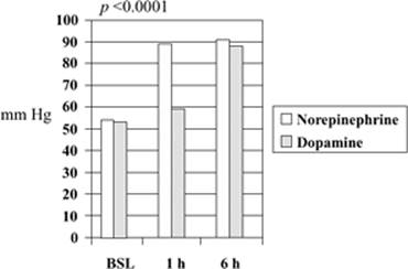
Figure 160.4. Histogram showing the effect of norepinephrine on mean arterial blood pressure (MAP) compared to high-dose dopamine in a randomized controlled trial in humans. MAP is more reliably restored using norepinephrine when given alone as an alternative to high-dose dopamine or after high-dose dopamine has failed. BSL, baseline. |
· Hypovolemia due to blood loss or third spacing
· Details of the operative field (e.g., aortic cross-clamping in major vascular surgery)
· Increases in intra-abdominal pressure (e.g., laparoscopic insufflation of CO2)
· Concomitant sepsis
· Use of nephrotoxin drugs
Any of these factors, alone or in combination, may contribute to a critical reduction in RBF and ischemia, impaired oxygen delivery, and toxin- or inflammatory-mediated injury. Postoperative ARF is believed to be, in part, mediated by proinflammatory mechanisms such as increased endothelial cell adhesion, tubular cell infiltration, generation of reactive oxygen species, proinflammatory cytokines, and reperfusion injury (51,52). Cardiac surgery with cardiopulmonary bypass (CPB) commonly induces early postoperative renal injury. The mechanisms whereby CPB causes injury are incompletely understood, although there is a suggestion that CPB is proinflammatory, activating components of the nonspecific immune system. In turn, this leads to oxidative stress with the generation of oxygen-free radical species and serum lipid peroxidation products (53). In addition, CPB has been shown to deplete serum antioxidative capacity for a prolonged duration after surgery. Such oxidant stress has been shown to directly induce renal injury in experimental studies (54).
ARF in Association with Mechanical Ventilation
Most critically ill patients require mechanical ventilation (MV), either for disease-specific indications such as acute respiratory distress syndrome (ARDS) or simply for routine postoperative care. The application of positive pressure MV, particularly with positive end-expiratory pressure (PEEP), can have important physiologic effects on kidney function. Experimental and clinical studies have clearly established an association between MV and PEEP and alterations in kidney function. This can occur through several mechanisms including (a) alterations in cardiovascular function, (b) alterations in neurohormonal activation, (c) abnormalities in gas exchange, and (d) alterations in systemic inflammatory mediators (55,56).
The positive pressure applied during MV acts to increase intrathoracic, intrapleural, and intra-abdominal pressures, both during inspiration and for the duration of the respiratory cycle. This increase in intrathoracic pressure, monitored clinically by changes in mean airway pressure, can act to reduce intrathoracic blood volume, decrease transmural pressure, reduce right ventricular preload, increase right ventricular afterload, exert alterations to pulmonary vascular resistance and volume, and contribute to changes in left ventricular filling and geometry. The result of these effects may be a decrease in cardiac output and renal perfusion. Similarly, raised intrathoracic pressure, by altering transmural pressures and reducing cardiac output, can act to unload intrathoracic baroreceptors. This initiates a cascade of compensatory neurohormonal events characterized by increased systemic and renal sympathetic nervous activity, increased activation of the renin–angiotensin–aldosterone system, increased secretion of vasopressin, and a reduction in release of atrial natriuretic peptide. These culminate in altered renal perfusion and kidney excretory function. Renal function may not be, per se, impaired with MV, but rather, may appropriately respond to stimuli by reducing osmolar, sodium, and water clearance. In addition, acute hypoxemia and/or hypercapnia, both commonly encountered in patients with ARDS, can act to alter systemic hemodynamics and increase systemic inflammation, both of which may exert negative effects on renal perfusion and function. Particular strategies of MV, specifically in ARDS, are now recognized to contribute to or provoke ventilator-induced lung injury (VILI). Evidence now suggests that the pathophysiology of VILI is multifactorial and results from the combined effects of volutrauma (excessive tidal or end-expiratory volumes), barotrauma (excessive end-inspiratory peak and plateau pressures), atelectatic trauma (cyclical opening and closing of alveolar units), and biotrauma (local release of inflammatory mediators from injured lung) (57). Such injurious MV can initiate a cascade of events that increase systemic inflammation and adversely affect kidney function (58).
The Clinical Picture
The most common clinical picture seen in the ICU is that of a patient who has sustained or is experiencing a major systemic insult such as trauma, sepsis, myocardial infarction, severe hemorrhage, cardiogenic shock, or major surgery. When the patient arrives in the ICU, resuscitation is typically well underway, or surgery may have just been completed. Despite such efforts, the patient is already anuric or profoundly oliguric, and the serum creatinine is rising, and a metabolic acidosis is developing; serum potassium and phosphate levels may be rapidly rising as well. In these critically ill patients with ARF, multiple organ dysfunction—with the need for mechanical ventilation and vasoactive drugs—is common. Fluid resuscitation is typically undertaken in the ICU with the guidance of invasive hemodynamic monitoring. Vasoactive drugs are often used to restore mean arterial pressure (MAP) to acceptable levels, typically greater than 65 to 70 mm Hg (Fig. 160.4). The patient may improve over time, and urine output may return with or without the assistance of diuretic agents (Fig. 160.5). If urine output does not return, however, renal replacement therapy (RRT) needs to be considered. If the cause of ARF has been removed, and the patient has become physiologically stable, slow recovery occurs within 4 to 5 days to as long as 3 or 4 weeks. In some cases, urine output can be above normal for several days. If the cause of ARF has not been adequately remedied, the patient remains gravely ill, the kidneys do not recover, and death from multiorgan failure may occur.
Preventing Arf
The fundamental principle of ARF prevention is to treat its cause. If prerenal factors contribute, these must be identified and hemodynamic resuscitation quickly instituted.
Fluid Resuscitation
Intravascular volume must be maintained or rapidly restored; this is often best done using invasive hemodynamic monitoring, such as with an arterial cannula and central venous catheter, pulmonary artery catheter, or pulse contour cardiac output catheter. Oxygenation must be maintained. An adequate hemoglobin concentration, usually at least more than about 7.0 g/dL, must be maintained or immediately restored. Once intravascular volume has been restored, some patients remain with a MAP less than 70 mm Hg. In these patients, autoregulation of RBF may be lost, and restoration of MAP to near normal levels may increase GFR (59,60,61). Such elevations in MAP, however, require the addition of vasopressor drugs (59,60,61). In patients with pre-existing hypertension or renovascular disease, a MAP of 75 to 80 mm Hg may still be inadequate. Experimental evidence suggests that vasopressor support in hypotensive sepsis increases renal blood flow (Fig. 160.6) and renal medullary blood flow (Fig. 160.7). The renal protective role of additional fluid therapy in a patient with a normal or increased cardiac output and blood pressure is questionable. Despite these resuscitation measures, renal failure may still develop if cardiac output is inadequate. This may require various interventions, from the use of inotropic drugs to the application of ventricular assist devices.
|
|
|
Figure 160.5. Diagram showing the effect of norepinephrine on urine output compared to high-dose dopamine in patients in septic shock. Urine output is more effectively restored with norepinephrine infusion when given alone as an alternative to high-dose dopamine or after high-dose dopamine has failed. BSL, baseline. |
Fluid Therapy
Fluid therapy is the cornerstone in resuscitation of the critically ill patient, and is the primary strategy for preservation of kidney function in the setting of increases in serum creatinine and/or urea, and oliguria. However, evolving evidence has suggested there may be negative consequences to overly aggressive fluid therapy for both renal and nonrenal organ function.
|
|
|
Figure 160.6. Diagram showing the changes in renal blood flow (RBF) during experimental E. coli–induced septic shock in sheep. The addition of norepinephrine increased renal blood flow. |
A large multicenter study found no significant difference in the incidence of ARF when comparing fluid resuscitation with crystalloid to albumin in critically ill patients (62). However, some synthetic colloid therapies, such as with the use of hydroxyethyl starches, have been associated with higher rates of ARF in critically ill patients after resuscitation for severe sepsis (63). Although the exact mechanism(s) remain uncertain, the hydroxyethyl starches may influence intrarenal hemodynamics or glomerular filtration through alterations in vascular oncotic pressure.
In critically ill patients, once apparent optimization of hemodynamics and intravascular volume status has been achieved, there is little evidence to support continued aggressive fluid resuscitation to improve kidney function (64). Rather, there is evidence from recent studies to suggest that such continued fluid administration and a positive cumulative balance can contribute to notable deteriorations in nonrenal organ function, in particular that of the lung (65,66). The ARDS Clinical Trials Network has completed the largest randomized trial assessing fluid therapy in patients with lung injury (67). This trial compared restrictive and liberal strategies for fluid management in 1,000 critically ill patients, mostly in those with pneumonia or sepsis with evidence of acute lung injury. At 72 hours, those receiving a restrictive fluid strategy had a near neutral fluid balance, whereas those in the liberal strategy were positive with more than 5 L. Although the study failed to show a difference in mortality between the strategies, a restrictive strategy improved lung function, increased ventilator-free days, and reduced ICU length of stay. Moreover, those in the restrictive group had a trend toward a reduced need for RRT.
|
|
|
Figure 160.7. Diagram showing the changes in medullary renal blood flow during experimental septic shock in sheep induced by E. coli administration. The addition of norepinephrine increased medullary blood flow. |
Renal Protective Drugs
Following hemodynamic resuscitation and removal of nephrotoxins, it is unclear whether the use of additional pharmacologic measures is of further benefit to the kidneys.
Renal Dose or Low-Dose Dopamine
Evidence of the efficacy or safety of the administration of dopamine in critically ill patients is lacking. However, this agent is a tubular diuretic and occasionally increases urine output. This may be incorrectly interpreted as an increase in GFR. Furthermore, a recent large phase III trial in critically ill patients showed low-dose dopamine to be as effective as placebo in the prevention of renal dysfunction (68) (Fig. 160.8).
Mannitol
A biologic rationale exists for the use of mannitol, as is the case for dopamine. However, no controlled human data exist to support its clinical use. The effect of mannitol as a renal protective agent remains questionable.
Loop Diuretics
These agents may protect the loop of Henle from ischemia by decreasing its transport-related workload. Animal data are encouraging, as are ex vivo experiments. There are no double-blind randomized controlled studies of suitable size to prove that these agents reduce the incidence of renal failure. However, some studies support the view that loop diuretics may decrease the need for RRT in patients developing ARF (69). They appear to achieve this by inducing polyuria, which allows for easier control of volume overload, acidosis, and hyperkalemia, the three major triggers for RRT in the ICU. Because avoiding dialysis simplifies treatment and reduces the cost of care, loop diuretics are occasionally used in patients with renal dysfunction, especially in the form of continuous infusion.
|
|
|
Figure 160.8. Diagram showing the comparative effects of placebo and renal-dose dopamine on peak serum creatinine and change in serum creatinine in critically ill patients from a large double-blinded randomized controlled trial. Renal-dose dopamine had no effect on serum creatinine. |
Other Agents
Other agents such as theophylline, urodilatin, and anaritide, a synthetic atrial natriuretic factor, have also been proposed. Studies so far, however, have been either experimental or underpowered, or have shown no beneficial effect. In a randomized double-blind, placebo-controlled trial, fenoldopam was shown to attenuate the deterioration in serum creatinine typically seen in septic patients (70). Studies of fenoldopam in other situations, however, have failed to show similar benefit (71). Thus, its role in ARF remains uncertain. Similarly, in a single-center study, recombinant human atrial natriuretic factor (rhANF) has been shown to attenuate renal injury in higher-risk patients undergoing cardiac surgery (72), but a large multicenter study of ARF failed to show a benefit (73). Many more investigations are urgently needed in this field.
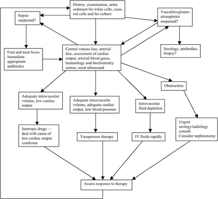
Diagnostic Investigations
An etiologic diagnosis of ARF must always be established (Fig. 160.9). Although such diagnosis may be obvious on clinical grounds, in many patients it is best to consider all possibilities and exclude common treatable causes by simple investigations. One such investigation includes microscopic examination of the urinary sediment. Urinalysis is a simple and noninvasive test that yields important diagnostic information and patterns suggestive of specific syndromes. The finding of dysmorphic red blood cells (RBC) or RBC casts is virtually diagnostic of active glomerulonephritis or vasculitis. Heavy proteinuria suggests some form of glomerular disease. White blood cell casts can suggest either interstitial nephropathy or infection. Similarly, a normal urinalysis can provide important information and can suggest that ARF is due to a prerenal or obstructive cause. Finally, examination of urine will provide evidence of whether a urinary tract infection is present.
|
|
|
Figure 160.9. Diagnostic and treatment approach to an intensive care unit patient presenting with acute renal failure. |
Several additional investigations may be necessary to establish the diagnosis. Evidence of marked anemia in the absence of blood loss may suggest acute hemolysis, thrombotic microangiopathy, or paraproteinemia related to malignancy. In microangiopathic hemolytic anemia, a peripheral blood smear will typically show evidence of hemolysis with the presence of schistocytes; the additional measurement of lactic dehydrogenase, haptoglobin, unconjugated bilirubin, and free hemoglobin are needed. If paraproteinemia due to multiple myeloma or lymphoma is suspected, serum and urine protein electrophoresis and serum calcium should be measured. A history of recent cancer diagnosis or chemotherapy should prompt the measurement of uric acid for tumor lysis syndrome.
In patients with a possible mechanism for muscle injury, creatine kinase and free myoglobin for possible rhabdomyolysis should be determined. If an elevated anion gap metabolic acidosis is present with suggestion of a toxic ingestion, ethylene glycol, methanol, and salicylates should be measured.
Systemic eosinophilia may be a clue suggesting systemic vasculitis, allergic interstitial nephritis, or atheroembolic disease. The measurement of specific antibodies—antiglomerular basement membrane (GBM), antineutrophil cytoplasmic antibodies (ANCA), antinuclear antibodies (ANA), anti-DNA, antismooth muscle, and so forth—or cryoglobulins are extremely useful screening tests to support the diagnosis of vasculitis or certain types of collagen vascular diseases or glomerulonephritis.
Imaging by renal ultrasonography is a rapid noninvasive investigation principally designed to rule out evidence of obstruction, stones, cysts, masses, or overt renovascular disease.
A chest radiograph may be important both to assess for pulmonary complications of ARF and if a diagnosis of systemic vasculitis is considered. In the occasional patient, a percutaneous renal biopsy becomes necessary to confirm the diagnosis, determine the severity of renal injury, guide therapy, and estimate the potential for renal recovery (74). A renal biopsy is indicated when a thorough noninvasive investigation has failed to yield the diagnosis, when prerenal and postrenal causes have been excluded, and prior to the administration of aggressive immunosuppressive therapy. A renal biopsy can be performed under ultrasound guidance with local anesthetic in critically ill patients undergoing mechanical ventilation without additional risks when compared to standard conditions.
Management of Established Acute Renal Failure
The principles of management of established ARF should always include the following:
· Confirmation of probable cause
· Elimination of potential contributors
· Institution of disease-specific therapy if applicable
· Prevention and management of ARF complications with maintenance of physiologic homeostasis while recovery takes place
Complications such as encephalopathy, pericarditis, myopathy, neuropathy, electrolyte disturbances, or other major electrolyte, fluid, or metabolic derangements should never occur in a modern ICU. They can be prevented by several measures, which vary in complexity from fluid restriction to the initiation of extracorporeal RRT.
Nutritional support must be started early and must contain adequate calories, around 30 to 35 Kcal/kg/day, as a mixture of carbohydrates and lipids. Sufficient protein of at least 1 to 2 g/kg/day must be administered. There is no evidence that specific renal nutritional solutions are useful. Vitamins and trace elements should be administered at least according to their recommended daily allowance. The role of newer immunonutritional solution remains controversial. The enteral route is preferred to the use of parenteral nutrition.
Hyperkalemia—a serum potassium level of greater than 6 mmol/L—must be promptly treated either with insulin and dextrose administration, the infusion of bicarbonate if acidosis is present, the administration of nebulized salbutamol, or all of the above combined. If the “true” serum potassium is more than 7 mmol/L, or if electrocardiographic signs of hyperkalemia appear, calcium gluconate—10 mL of 10% solution administered IV—should also be used. The above measures are temporizing actions while RRT is being arranged. The presence of hyperkalemia is a major indication for the immediate institution of RRT.
Metabolic acidosis, almost always present, rarely requires treatment per se. Anemia requires correction to maintain a hemoglobin greater than about 7.0 g/dL; more aggressive transfusion may be needed based on individual patient assessment (75). Drug therapy must be adjusted to take into account the effect of the decreased clearances associated with loss of renal function. Stress ulcer prophylaxis is advisable and should be based on H2-receptor antagonists or proton pump inhibitors in selected cases. Assiduous attention should be paid to the prevention of infection.
|
Table 160.3 Interventions That May Potentially Influence Renal Recovery Renal Replacement Therapy (RRT) |
|
|
Fluid overload can be prevented by the use of loop diuretics in polyuric patients. However, if the patient is oliguric, the only way to avoid fluid overload is to institute RRT at an early stage. Marked azotemia, defined as a urea more than 40 mmol/L (BUN [blood urea nitrogen] of 112 mg/dL) or a creatinine level more than 400 micromoles (4.5 mg/dL), is undesirable and should probably be treated with RRT, unless recovery is imminent or already under way and a return toward normal values is expected within 24 to 48 hours. It is recognized, however, that no randomized trials exist to define the ideal time for intervention with artificial renal support.
Recovery and Its Management
Recovery of renal function after an episode of ARF is increasingly being acknowledged as a significant clinical measure of morbidity. Failure to recover function can have both individual patient and broader health care implications. Persistent chronic renal impairment, or the need for long-term RRT, can negatively influence the health status and quality of life of patients and contribute to considerable annual health care expenditures. Recovery to independence from RRT occurs in an estimated 68% to 85% of critically ill patients by hospital discharge and generally peaks by 90 days (1,4). Studies have shown that older patients and those with pre-existing comorbid illnesses, such as chronic kidney disease or advanced cardiovascular disease, are less likely to recover function, whereas those with septic ARF may be more likely to recover function. Several other potentially modifiable factors have been linked with improved rates of recovery, including early and aggressive initiation of RRT when indicated, use of continuous rather than intermittent RRT, early and adequate nutritional support, and intensive insulin therapy (Table 160.3). Whether adjuvant erythropoietin and routine use of loop diuretics can influence renal prognosis and promote early recovery remain controversial.
Prognosis
Acute renal failure can independently influence both short- and long-term prognosis. In hospitalized patients, mortality is estimated at 20% among all those developing ARF; however, this rate is greatly influenced by the severity of renal injury. The prognosis is worse for critically ill patients and those in whom RRT becomes necessary. The in-hospital mortality for critically ill patients with ARF is estimated at 50% to 60%, yet, depending on the case mix, can range between 40% to 80% (1,3,4,76,77). It is frequently stated that patients die with renal failure rather than of renal failure. However, growing evidence suggests that better uremic control and more intensive artificial renal support may improve survival (78,79). Such evidence supports a careful and proactive approach to the treatment of critically ill patients with ARF, which is based on the prevention of uncontrolled uremia and the maintenance of low urea levels throughout the patient's illness.
In those who survive an episode of ARF associated with critical illness, the long-term health status, including health-related quality of life (HRQoL), functional status, and hospital discharge location, are also now considered important indicators of morbidity. These patients frequently describe limitations in daily activities, defficulties with mobility, and high levels of sleep disturbance, fatigue, anxiety, and depression. However, HRQoL is generally good and perceived as acceptable, despite evidence that their quality of life is considerably lower than that of the general population (80).
Future Developments
The discipline of nephrology concerned with ARF and RRT in critically ill patients has undergone remarkable progress in recent years; however, mortality rates for ARF remain unacceptably high. A consensus definition has now been developed and published that will guide research and, it is hoped, translate into improved patient outcome (6). Such research is needed to explore the relationship between survival and subsequent morbidity—specifically, recovery of kidney function, health-related quality of life, and the economic consequences of decisions made during care of critically ill patients with ARF.
Some recent advances have been made, particularly in the prevention of ARF associated with radiocontrast nephropathy with use of N-acetylcysteine. Although studies with fenoldopam are provocative, in general, no specific drugs have been found to help. Apoptosis has recently been shown as an important mechanism of renal tubular injury in ARF (47,48,49,86). There is therapeutic potential for molecular targets, such as selective inhibitors of pro-apoptotic proteins (e.g., capsase inhibitors), involved in ARF that may attenuate injury or promote recovery; however, at present, no evidence in humans has emerged.
Pearls
· Restoring mean arterial pressure (MAP) within the autoregulatory range for blood flow to the kidney—65 to 110 mm Hg—is important in maintaining the glomerular filtration rate. Once the patient has been adequately fluid resuscitated, with a CVP at least greater than 8 mm Hg, and the cardiac output is known to be adequate or high, MAP should be corrected within autoregulation with the use of norepinephrine.
· Low-dose dopamine has been extensively studied, meta-analyzed, and assessed for the treatment of acute renal failure. Although it probably increases urine output through its tubular diuretic effect, it does not maintain or improve the glomerular filtration rate.
· Once a patient has been fluid resuscitated as described above, if the cardiac output is adequate or high, and the mean arterial pressure is adequate or normal, there is no renal benefit to be gained by giving more intravenous fluids. Such fluids often precipitate pulmonary congestion and have no sustained beneficial effect on glomerular filtration. They should not be given.
References
1. Bagshaw SM, Laupland KB, Doig CJ, et al. Prognosis for long-term survival and renal recovery in critically ill patients with severe acute renal failure: a population-based study. Crit Care. 2005;9(6):R700–R709.
2. de Mendonca A, Vincent JL, Suter PM, et al. Acute renal failure in the ICU: risk factors and outcome evaluated by the SOFA score. Intensive Care Med. 2000;26(7):915–921.
3. Liano F, Junco E, Pascual J, et al. The spectrum of acute renal failure in the intensive care unit compared with that seen in other settings. The Madrid Acute Renal Failure Study Group. Kidney Int Suppl. 1998;66:S16–24.
4. Uchino S, Kellum JA, Bellomo R, et al. Acute renal failure in critically ill patients: a multinational, multicenter study. JAMA. 2005;294(7):813–818.
5. Metnitz PG, Krenn CG, Steltzer H, et al. Effect of acute renal failure requiring renal replacement therapy on outcome in critically ill patients. Crit Care Med. 2002;30(9):2051–2058.
6. Bellomo R, Ronco C, Kellum JA, et al. Acute renal failure—definition, outcome measures, animal models, fluid therapy and information technology needs: the Second International Consensus Conference of the Acute Dialysis Quality Initiative (ADQI) Group. Crit Care. 2004;8(4):R204–212.
7. Uchino S, Bellomo R, Goldsmith D, et al. An assessment of the RIFLE criteria for acute renal failure in hospitalized patients. Crit Care Med. 2006;34:1913–1917.
8. Hoste EA, Clermont G, Kersten A, et al. RIFLE criteria for acute kidney injury are associated with hospital mortality in critically ill patients: a cohort analysis. Crit Care. 2006;10(3):R73.
9. Hou SH, Bushinsky DA, Wish JB, et al. Hospital-acquired renal insufficiency: a prospective study. Am J Med. 1983;74(2):243–248.
10. Nash K, Hafeez A, Hou S. Hospital-acquired renal insufficiency. Am J Kidney Dis. 2002;39(5):930–936.
11. McCarthy JT. Prognosis of patients with acute renal failure in the intensive-care unit: a tale of two eras. Mayo Clin Proc. 1996;71(2):117–126.
12. Tran DD, Cuesta MA, Oe PL. Acute renal failure in patients with severe civilian trauma. Nephrol Dial Transplant. 1994;9(Suppl 4):121–125.
13. Bagshaw SM, Langenberg C, Bellomo R. Urinary biochemistry and microscopy in septic acute renal failure—a systematic review. Am J Kidney Dis. 2006;48(5):695–705.
14. Langenberg C, Wan L, Bagshaw SM, et al. Urinary biochemistry in experimental septic acute renal failure. Nephrol Dial Transplant. 2006;21(12):3389–3397.
15. Feest T, Round A, Hamad S. Incidence of severe acute renal failure in adults: results of a community based study. BMJ. 1993;306:481–483.
16. Gines P, Guevara M, Arroyo V, et al. Hepatorenal syndrome. Lancet. 2003;362(9398):1819–1827.
17. Arroyo V, Guevara M, Gines P. Hepatorenal syndrome in cirrhosis: pathogenesis and treatment. Gastroenterology. 2002;122(6):1658–1676.
18. Sort P, Navasa M, Arroyo V, et al. Effect of intravenous albumin on renal impairment and mortality in patients with cirrhosis and spontaneous bacterial peritonitis. N Engl J Med. 1999;341(6):403–409.
19. Guevara M, Gines P, Fernandez-Esparrach G, et al. Reversibility of hepatorenal syndrome by prolonged administration of ornipressin and plasma volume expansion. Hepatology. 1998;27(1):35–41.
20. Fabrizi F, Dixit V, Martin P. Meta-analysis: terlipressin therapy for the hepatorenal syndrome. Aliment Pharmacol Ther. 2006;24(6):935–944.
21. Guevara M, Gines P, Bandi JC, et al. Transjugular intrahepatic portosystemic shunt in hepatorenal syndrome: effects on renal function and vasoactive systems. Hepatology. 1998;28(2):416–422.
22. Brensing KA, Textor J, Perz J, et al. Long term outcome after transjugular intrahepatic portosystemic stent-shunt in non-transplant cirrhotics with hepatorenal syndrome: a phase II study. Gut. 2000;47(2):288–295.
23. Silvester W, Bellomo R, Cole L. Epidemiology, management, and outcome of severe acute renal failure of critical illness in Australia. Crit Care Med. 2001;29(10):1910–1915.
24. Holt SG, Moore KP. Pathogenesis and treatment of renal dysfunction in rhabdomyolysis. Intensive Care Med. 2001;27(5):803–811.
25. Heyman SN, Brezis M, Reubinoff CA, et al. Acute renal failure with selective medullary injury in the rat. J Clin Invest. 1988;82(2):401–412.
26. Bonventre JV. Mechanisms of ischemic acute renal failure. Kidney Int. 1993;43(5):1160–1178.
27. di Mari JF, Davis R, Safirstein RL. MAPK activation determines renal epithelial cell survival during oxidative injury. Am J Physiol. 1999;277(2 Pt 2):F195–203.
28. Portilla D, Mandel LJ, Bar-Sagi D, Millington DS. Anoxia induces phospholipase A2 activation in rabbit renal proximal tubules. Am J Physiol. 1992;262(3 Pt 2):F354–360.
29. Bennett WM, Luft F, Porter GA. Pathogenesis of renal failure due to aminoglycosides and contrast media used in roentgenography. Am J Med. 1980;69(5):767–774.
30. Cunha MA, Schor N. Effects of gentamicin, lipopolysaccharide, and contrast media on immortalized proximal tubular cells. Ren Fail. 2002;24(6):687–690.
31. Bagshaw SM, Culleton BF. Contrast-induced nephropathy: epidemiology and prevention. Minerva Cardioangiol. 2006;54(1):109–129.
32. Aspelin P, Aubry P, Fransson SG, et al. Nephrotoxic effects in high-risk patients undergoing angiography. N Engl J Med. 2003;348(6):491–499.
33. Merten GJ, Burgess WP, Gray LV, et al. Prevention of contrast-induced nephropathy with sodium bicarbonate: a randomized controlled trial. JAMA. 2004;291(19):2328–2334.
34. Mueller C, Seidensticker P, Buettner HJ, et al. Incidence of contrast nephropathy in patients receiving comprehensive intravenous and oral hydration. Swiss Med Wkly. 2005;135(19–20):286–290.
35. Stevens MA, McCullough PA, Tobin KJ, et al. A prospective randomized trial of prevention measures in patients at high risk for contrast nephropathy: results of the P.R.I.N.C.E. Study. Prevention of Radiocontrast Induced Nephropathy Clinical Evaluation. J Am Coll Cardiol. 1999;33(2):403–411.
36. Bagshaw SM, Ghali WA. Acetylcysteine for prevention of contrast-induced nephropathy after intravascular angiography: a systematic review and meta-analysis. BMC Med. 2004;2:38.
37. Tepel M, van der Giet M, Schwarzfeld C, et al. Prevention of radiographic-contrast-agent-induced reductions in renal function by acetylcysteine. N Engl J Med. 2000;343(3):180–184.
38. Langenberg C, Bellomo R, May C, et al. Renal blood flow in sepsis. Crit Care. 2005;9(4):R363–3674.
39. Langenberg C, Bellomo R, May CN, et al. Renal vascular resistance in sepsis. Nephron Physiol. 2006;104(1):1–11.
40. Langenberg C, Wan L, Egi M, et al. Renal blood flow in experimental septic acute renal failure. Kidney Int. 2006;69(11):1996–2002.
41. Lucas CE, Rector FE, Werner M, et al. Altered renal homeostasis with acute sepsis. Clinical significance. Arch Surg. 1973;106(4):444–449.
42. Rector F, Goyal S, Rosenberg IK, et al. Renal hyperemia in associated with clinical sepsis. Surg Forum. 1972;23:51–53.
43. Brenner M, Schaer GL, Mallory DL, et al. Detection of renal blood flow abnormalities in septic and critically ill patients using a newly designed indwelling thermodilution renal vein catheter. Chest. 1990;98(1):170–179.
44. Di Giantomasso D, Morimatsu H, May CN, et al. Intrarenal blood flow distribution in hyperdynamic septic shock: effect of norepinephrine. Crit Care Med. 2003;31(10):2509–2513.
45. Hotchkiss RS, Swanson PE, Freeman BD, et al. Apoptotic cell death in patients with sepsis, shock, and multiple organ dysfunction. Crit Care Med. 1999;27(7):1230–1251.
46. Wan L, Bellomo R, Di Giantomasso D, et al. The pathogenesis of septic acute renal failure. Curr Opin Crit Care. 2003;9(6):496–502.
47. Jo SK, Cha DR, Cho WY, et al. Inflammatory cytokines and lipopolysaccharide induce Fas-mediated apoptosis in renal tubular cells. Nephron. 2002;91(3):406–415.
48. Messmer UK, Briner VA, Pfeilschifter J. Tumor necrosis factor-alpha and lipopolysaccharide induce apoptotic cell death in bovine glomerular endothelial cells. Kidney Int. 1999;55(6):2322–2337.
49. Bonegio R, Lieberthal W. Role of apoptosis in the pathogenesis of acute renal failure. Curr Opin Nephrol Hypertens. 2002;11(3):301–308.
50. Imai Y, Parodo J, Kajikawa O, et al. Injurious mechanical ventilation and end-organ epithelial cell apoptosis and organ dysfunction in an experimental model of acute respiratory distress syndrome. JAMA. 2003;289(16):2104–2112.
51. Gueler F, Rong S, Park JK, et al. Postischemic acute renal failure is reduced by short-term statin treatment in a rat model. J Am Soc Nephrol. 2002;13(9):2288–2298.
52. Noiri E, Nakao A, Uchida K, et al. Oxidative and nitrosative stress in acute renal ischemia. Am J Physiol Renal Physiol. 2001;281(5):F948–957.
53. Starkopf J, Zilmer K, Vihalemm T, et al. Time course of oxidative stress during open-heart surgery. Scand J Thorac Cardiovasc Surg. 1995;29(4):181–186.
54. Ishizuka S, Nagashima Y, Numata M, et al. Regulation and immunohistochemical analysis of stress protein heme oxygenase-1 in rat kidney with myoglobinuric acute renal failure. Biochem Biophys Res Commun. 1997;240(1):93–98.
55. Kuiper JW, Groeneveld AB, Slutsky AS, et al. Mechanical ventilation and acute renal failure. Crit Care Med. 2005;33(6):1408–1415.
56. Pannu N, Mehta RL. Mechanical ventilation and renal function: an area for concern? Am J Kidney Dis. 2002;39(3):616–624.
57. Ricard JD, Dreyfuss D, Saumon G. Ventilator-induced lung injury. Curr Opin Crit Care. 2002;8(1):12–20.
58. Ranieri VM, Suter PM, Tortorella C, et al. Effect of mechanical ventilation on inflammatory mediators in patients with acute respiratory distress syndrome: a randomized controlled trial. JAMA. 1999;282(1):54–61.
59. Albanese J, Leone M, Garnier F, et al. Renal effects of norepinephrine in septic and nonseptic patients. Chest. 2004;126(2):534–539.
60. Bellomo R, Kellum JA, Wisniewski SR, et al. Effects of norepinephrine on the renal vasculature in normal and endotoxemic dogs. Am J Respir Crit Care Med. 1999;159(4 Pt 1):1186–1192.
61. Bourgoin A, Leone M, Delmas A, et al. Increasing mean arterial pressure in patients with septic shock: effects on oxygen variables and renal function. Crit Care Med. 2005;33(4):780–786.
62. Finfer S, Bellomo R, Boyce N, et al. A comparison of albumin and saline for fluid resuscitation in the intensive care unit. N Engl J Med. 2004;350(22):2247–2256.
63. Schortgen F, Lacherade JC, Bruneel F, et al. Effects of hydroxyethylstarch and gelatin on renal function in severe sepsis: a multicentre randomised study. Lancet. 2001;357(9260):911–916.
64. Van Biesen W, Yegenaga I, Vanholder R, et al. Relationship between fluid status and its management on acute renal failure (ARF) in intensive care unit (ICU) patients with sepsis: a prospective analysis. J Nephrol. 2005;18(1):54–60.
65. Sakr Y, Vincent JL, Reinhart K, et al. High tidal volume and positive fluid balance are associated with worse outcome in acute lung injury. Chest. 2005;128(5):3098–3108.
66. Simmons RS, Berdine GG, Seidenfeld JJ, et al. Fluid balance and the adult respiratory distress syndrome. Am Rev Respir Dis. 1987;135(4):924–929.
67. Wiedemann HP, Wheeler AP, Bernard GR, et al. Comparison of two fluid-management strategies in acute lung injury. N Engl J Med. 2006;354(24):2564–2575.
68. Bellomo R, Chapman M, Finfer S, et al. Low-dose dopamine in patients with early renal dysfunction: a placebo-controlled randomised trial. Australian and New Zealand Intensive Care Society (ANZICS) Clinical Trials Group. Lancet. 2000;356(9248):2139–2143.
69. Shilliday IR, Quinn KJ, Allison ME. Loop diuretics in the management of acute renal failure: a prospective, double-blind, placebo-controlled, randomized study. Nephrol Dial Transpl. 1997;12(12):2592–2596.
70. Morelli A, Ricci Z, Bellomo R, et al. Prophylactic fenoldopam for renal protection in sepsis: a randomized, double-blind, placebo-controlled pilot trial. Crit Care Med. 2005;33(11):2451–2456.
71. Bove T, Landoni G, Calabro MG, et al. Renoprotective action of fenoldopam in high-risk patients undergoing cardiac surgery: a prospective, double-blind, randomized clinical trial. Circulation. 2005;111(24):3230–3235.
72. Sward K, Valsson F, Odencrants P, et al. Recombinant human atrial natriuretic peptide in ischemic acute renal failure: a randomized placebo-controlled trial. Crit Care Med. 2004;32(6):1310–1315.
73. Chertow GM, Lazarus JM, Paganini EP, et al. Predictors of mortality and the provision of dialysis in patients with acute tubular necrosis. The Auriculin Anaritide Acute Renal Failure Study Group. J Am Soc Nephrol. 1998;9(4):692–698.
74. Korbet SM. Percutaneous renal biopsy. Semin Nephrol. 2002;22(3):254–267.
75. Hebert PC, Wells G, Blajchman MA, et al. A multicenter, randomized, controlled clinical trial of transfusion requirements in critical care. Transfusion Requirements in Critical Care Investigators, Canadian Critical Care Trials Group. N Engl J Med. 1999;340(6):409–417.
76. Mehta RL, Pascual MT, Soroko S, et al. Spectrum of acute renal failure in the intensive care unit: the PICARD experience. Kidney Int. 2004;66(4):1613–1621.
77. Ympa YP, Sakr Y, Reinhart K, et al. Has mortality from acute renal failure decreased? A systematic review of the literature. Am J Med. 2005;118(8):827–832.
78. Schiffl H, Lang SM, Fischer R. Daily hemodialysis and the outcome of acute renal failure. N Engl J Med 2002;346:305–310.
79. Phu NH, Hien TT, Hoang NT, et al. Hemofiltration and peritoneal dialysis in infection-associated acute renal failure in Vietnam. N Engl J Med 2002;347:895–902.
80. Ronco C, Bellomo R, Homel P, et al. Effects of different doses in continuous veno-venous haemofiltration on outcomes of acute renal failure: a prospective randomised trail. Lancet 2000;355:26–30.