Q. If the tumour was an NSGCT but still stage I disease, would you do anything different compared to clinical stage I seminoma?
A. Again, a number of options exist post-radical orchidectomy with surveillance or adjuvant chemotherapy or, for those not prepared to consider the first two options, RPLND remains an option.
Across all NSGCTs surveillance alone has a 30% relapse rate (the majority in the retroperitoneum and then the lungs) if orchidectomy is the sole treatment. The majority of such relapses occur in the first year of follow-up (~80%).
Q. Is there anything that can guide you in determining and counselling the patient about the most appropriate option?
A. For NSGCTs the presence of vascular invasion is the most important prognosticator for distant relapse. The presence of vascular invasion portends a 48% risk of developing metastatic disease versus 14%-22% without vascular invasion. Once again, a risk-adapted approach can be used to stratify patients into low-risk and high-risk groups based on the absence or presence of vascular invasion, respectively.
Q. What would you advise the patient?
A. The patient can choose adjuvant treatment but in the absence of vascular invasion we would advocate a surveillance program with regular CT scans at 0, 3 and 12 months. This means that 78%-86% of patients are cured following orchidectomy alone and do not require further treatment.
However, if there are difficulties with patient compliance with this regime, adjuvant chemotherapy with two cycles of BEP are recommended.
However, in the presence of vascular invasion, we would advise adjuvant chemotherapy with two cycles of BEP. Surveillance can be used but given the 48% risk of relapse and the anxiety this can generate adjuvant chemotherapy is advised.
Q. Is there anything else that can be offered in clinical stage I NSGCT with vascular invasion in this group of patients?
A. Occasionally, patients unwilling to undergo surveillance or adjuvant chemotherapy can be offered an RPLND. This exposes patients to surgery and its associated side effects in about 50% of cases who may never have relapsed. At the same time RPLND does not eliminate the possibility of late distant recurrence, often in the lungs (~10%). In such patients who undergo an RPLND ~30% have disease in the retroperitoneum that upstages their disease to pathological stage II disease and will require additional chemotherapy with two cycles of BEP to reduce the 30% risk of relapse to 2%, although in some centres surgery alone will be used. In the United States it is normal practice to offer all stage I NSGCTs an RPLND to accurately pathologically stage the disease. In the United Kingdom, first-line treatment is surveillance or two cycles of BEP chemotherapy. If the patient is unwilling to follow an intensive surveillance program or undergo chemotherapy an RPLND can be advised.
Q. In general what would the follow-up be for these patients?
A. The follow-up schedule differs depending on the treatment modality chosen. For those who choose surveillance this is more intensive and regimens vary but generally patients require three-monthly clinic visits with tumour marker assessment, biannual chest x-rays and biannual abdomino-pelvic CT for the first 2 years with the frequency of these tailing off with up to 10 years of follow-up. For those who choose RPLND or adjuvant chemotherapy the follow-up is less intensive with the above applicable in general but with the CT scan being undertaken annually.
Q. If instead the patient at first presentation on his staging CT had the following appearances, Figure 2.5, with elevated tumour markers what would you do?
A. He has clinical stage II disease and I would There fore offer the patient induction chemotherapy based on the IGCCCG prognostic classification (Table 2.6). For the good prognosis group, they would normally receive three cycles of BEP (22 day cycle), or for those in whom bleomycin needs to be avoided, four cycles of EP. For the intermediate and poor groups, four cycles of BEP, or if bleomycin is to be avoided, then four cycles of cisplatin, etoposide and ifosfamide (PEI/VIP) with a follow-up repeat CT scan to look for regression of the retroperitoneal nodes at 4-6 weeks. Occasionally, a rare situation arises where small retroperitoneal nodes are detected, that may well be benign, without elevation of tumour markers (marker negative stage IIA disease). The difficulty is deciding whether such nodes are pathological. For marker negative stage IIA disease with small 1-2 cm nodes, a policy of surveillance especially if the nodes are shrinking on serial scans is acceptable although this depends also on the original orchidectomy histology. The EAU recommends that in clinical stage IIA disease with negative markers, patients with pure embryonal carcinoma in the orchidectomy specimen undergo immediate chemotherapy.
If the histology was teratoma or mixed tumour, surveillance or an RPLND can be undertaken. In practice in the United Kingdom, most centres would give chemotherapy or an early CT and if the nodes were still present, proceed with chemotherapy. Patients with marker negative stage IIA disease who are unwilling to undergo chemotherapy have the option to have RPLND with adjuvant chemotherapy (two cycles of BEP) if nodal disease is present on RPLND histology.

Figure 2.5
Table 2.6 The International Germ Cell Cancer Collaborative Group (IGCCCG) prognostic-based staging system for metastatic NSGCT
|
Good prognosis group |
|
|
(56% of cases) |
All of the following criteria |
|
5-year PFS 89% |
Testis/retroperitoneal primary |
|
5-year survival 92% |
No non-pulmonary visceral metastases |
|
AFP < 1000 ng/mL |
|
|
hCG < 5000 IU/L (1,000 ng/mL) |
|
|
LDHz < 1.5 x ULN |
|
|
Intermediate prognosis group |
|
|
(28% of cases) |
Any of the following criteria |
|
5-year PFS 41% |
Mediastinal primary |
|
5-year survival 48% |
Non-pulmonary visceral metastases |
|
AFP > 1000 and < 10,000 ng/mL or |
|
|
hCG > 5000 and <50,000 IU/L or |
|
|
LDH > 1.5 and <10 x ULN |
|
|
Poor prognosis group |
|
|
(16% of cases) |
Any of the following criteria |
|
5-year PFS 41% |
Mediastinal primary |
|
5-year survival 48% |
Non-pulmonary visceral metastases |
|
AFP > 10,000 ng/mL or |
|
|
hCG > 50,000 IU/L (10,000 ng/mL) or |
|
|
LDH > 10 x ULN |
|
Note: PFS - progression-free survival.
Q. His CT after induction chemotherapy is as shown in Figure 2.6. What would you do now?
A. There still remains a large para-aortic retroperitoneal mass that has regressed considerably following the chemotherapy. At this point I would check his tumour markers again. If these have normalised he has potentially resectable disease by way of an RPLND. However, if his markers remained elevated but at a plateau we would watch this a little further with further markers taken at variable times in the succeeding 4-12 weeks. If at this stage these remained stable we would offer him an RPLND. If they continue to rise then instead of surgery, salvage chemotherapy is needed with PEI/VIP or paclitaxel, ifosfamide and cisplatin (TIP) regimes.
ttere is no reliable model to predict whether such masses harbour active tumour and so RPLND is mandatory with residual masses in excess of 1 cm as the likelihood of significant residual disease rises. Unlike stage II seminoma there is no role for PET scanning in this group. In general, for those patients who undergo RPLND for a residual mass after primary induction BEP chemotherapy, the histology will show necrosis in 50% (~30% in most series), mature teratoma in 35% and viable cancer in 15%, although there are few predictors of this. Such masses have an increased risk of harbouring teratoma in the final histology if the original orchidectomy specimen had teratoma.

Figure 2.6
Q. A different patient had initially presented with clinical stage I NSGCT disease with vascular invasion and completed two cycles of primary BEP chemotherapy. He has a follow-up CT scan at 12 months. What do the images show - Figures 2.7 and 2.8?
A. Axial (Figure 2.7) and coronal (Figure 2.8) CT images show bulky nodes in the retroperitoneum encasing the great vessels indicating clinical relapse.
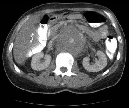
Figure 2.7

Figure 2.8
Q. What do you do now with the patient?
A. In this instance given a residual mass following induction chemotherapy, the patient needs second-line or salvage chemotherapy. This normally takes the form of four cycles of PEI/VIP (cisplatin, etoposide and ifosfamide) or four cycles of TIP (cisplatin, ifosfamide and paclitaxel). The response to this salvage chemotherapy depends on a variety of factors such as the original histology and location of the tumour, response to first-line treatment, duration of remission and level of tumour markers at relapse. There is some early evidence that treatment of refractory germ cell tumours may benefit from a combination of Taxol and gemcitabine chemotherapy and referral to centres that have expertise in this area as well as entering such patients into clinical trials is advised.
Q. If the retroperitoneal mass persisted despite salvage chemotherapy (Figure 2.9) is there anything else that could be offered?
A. Yes, salvage RPLND 4-6 weeks after normalisation of tumour markers or achievement of their plateau could be offered. The outlook is poor for those patients who after second- or third-line chemotherapy still harbour undifferentiated tumour in the surgical specimen.

Figure 2.9 Persistent retroperitoneal mass despite salvage chemotherapy.
Q. What are the principles of post-chemotherapy RPLND surgery?
A. Through a transabdominal or thoracoabdominal approach, the retroperitoneal great vessels are completely cleared removing all lymphatic tissue out to the ureters, extending from the renal artery down to the ipsilateral external iliac vessels. Nodal tissue is dissected out in an attempt to cure the patient (if this is the only site of metastatic disease) but one must minimise morbidity and particularly attempt to preserve antegrade ejaculation, if possible. With unilateral disease it may be possible to preserve the contralateral hypogastric plexus and postganglionic sympathetic fibres (and so antegrade ejaculation). In pre-chemotherapy cases nerve-sparing RPLND can be performed leading to an antegrade ejaculation rate as high as 90%.
Q. What would you warn the patient about prior to an RPLND?
A. RPLND is a major undertaking that has an associated mortality (1%-3%) and morbidity rate (5%-25%) and with the use of modern dissection templates is designed to minimise the complications mainly associated with ejaculation and subsequent fertility in young men. The main complications are as follows:
Overall complication rate ~10%
Major complications ~1%-5%
Chylous ascites - 1%-3%
Renovascular injury and nephrectomy - higher in post-chemotherapy cases - it is not ‘injury’ but a planned nephrectomy in around 5%-8%
Small bowel obstruction - 1%-3%
Spinal cord ischaemia - <1%
Minor complications ~15%
Wound infections
Anejaculation
Paralytic ileus
Lymphocele
Transient hyperamylasemia
Pneumonitis/atelectasis
Q. How would you follow-up a patient after an RPLND?
A. This depends on the histology. For necrosis and mature teratoma no further adjuvant treatment is required as the relapse rate in this group of patients is low (~5%-10%) but for
viable tumour further salvage chemotherapy is advised with second-line chemotherapy [21]. In completely resected tumours with viable tumour, 70% of patients remain disease free as compared to none who did not receive further chemotherapy. Furthermore, patients with complete resection, good IGCCCG prognostic grouping and less than 10% viable tumour, tend to fair well following RPLND and There fore may not need further salvage chemotherapy.
With respect to chemotherapy, caution is used in those who have already received bleomycin as this cumulative dose increases the risk of ‘bleomycin lung’, a pneumonitis and fibrotic condition of the lung interstitium. Occasionally if a further residual mass persists despite this, a second RPLND can be considered but this is rare.
Q. If the patient had first presented with stage III NSGCT disease how would the treatment broadly speaking differ?
A. Again the treatment would be similar to advanced seminoma based on the IGCCCG classification system. In essence this is three cycles of BEP chemotherapy in the first instance for the good prognosis group and four cycles for the intermediate group. For the poor prognostic group the standard remains four cycles of BEP or PEI (cisplatin, etoposide and ifosfamide) but referral to a reference centre for inclusion into trials is recommended as the optimum treatment remains to be established.
Q. If a patient at first presentation has a CT of the brain that shows a metastatic deposit, does that confer a poor prognosis compared to a later relapse to the brain following initial successful treatment (Figure 2.10)?
A. No. Approximately 10% of all patients with a germ cell tumour present with brain metastases and overall have a long-term survival of 30%-40%. Patients who relapse in the brain following initial treatment do so as part of a systemic relapse pattern and have a poor 5-year survival in the order of 2%-5%.

Figure 2.10